Does Skin Cancer Look Like A Scab
4.6/5doWhat it looks likeskinmore on it
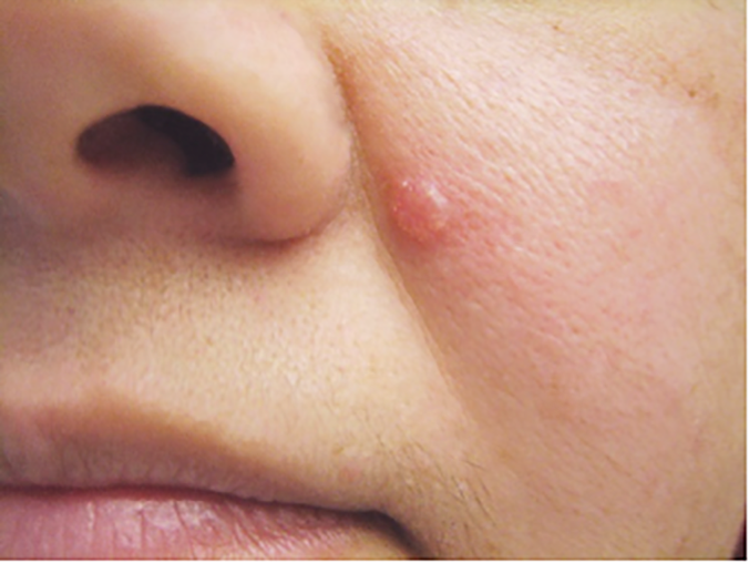
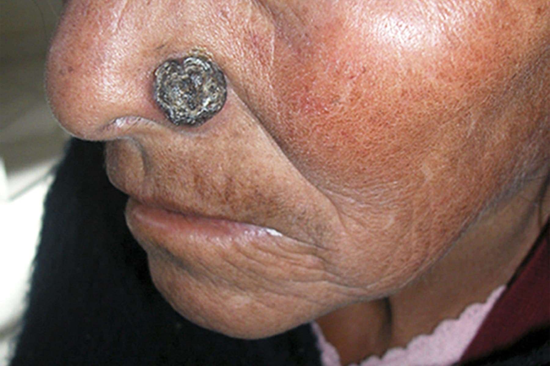
“Squamous cell cancers, which can metastasize if left untreated, are often reddish marks that will scab, flake off, then scab again,” Bank says. If you draw a line through the middle of a benign mole, the two halves will line up. Cancerous cells don’t grow evenly.
Furthermore, what does early signs of skin cancer look like? Melanoma signs include: A large brownish spot with darker speckles. A mole that changes in color, size or feel or that bleeds. A small lesion with an irregular border and portions that appear red, pink, white, blue or blue-black.
Also, does skin cancer scab and bleed?
The skin features that frequently develop are listed below. For basal cell carcinoma, 2 or more of the following features may be present: An open sore that bleeds, oozes, or crusts and remains open for several weeks. A reddish, raised patch or irritated area that may crust or itch, but rarely hurts.
What is a scab that won’t heal?
Chronic wounds, by definition, are sores that don’t heal within about three months. They can start small, as a pimple or a scratch. They might scab over again and again, but they don’t get better.
Ask The Expert: Why Am I Having Surgery To Remove A Small Basal Cell Carcinoma
Although the nonmelanoma skin cancer basal cell carcinoma is rarely life-threatening, it can be troublesome, especially because 80 percent of BCCs develop on highly visible areas of the head and neck. These BCCs can have a substantial impact on a persons appearance and can even cause significant disfigurement if not treated appropriately in a timely manner.
The fact is, BCCs can appear much smaller than they are. On critical areas of the face such as the eyes, nose, ears and lips, they are more likely to grow irregularly and extensively under the skins surface, and the surgery will have a greater impact on appearance than might have been guessed. Even a small BCC on the face can be deceptively large and deep the extent of the cancer cannot be seen with the naked eye.
If such a BCC is treated nonsurgically , the chance of the cancer recurring is high. Unfortunately, treating a BCC that has returned is usually much more difficult than treating it precisely and completely when initially diagnosed.
BCCs on the trunk, arms and legs that cause concern are typically larger in size, but even a small BCC in these areas can have an irregular growth pattern under the skin if the initial biopsy shows the tumor is aggressive. In addition, a small BCC in an area previously treated with radiation may be much more aggressive than it appears on the surface. Again, treating such a tumor nonsurgically is likely to leave cancer cells behind.
About the Expert:
Painful Skin Lesions May Signal Squamous Cell Carcinoma
When dermatology patients report painful skin lesions, their doctors may want to look for squamous cell carcinoma, according to the latest research findings from Wake Forest Baptist Medical Center.
Gil Yosipovitch, M.D., professor of dermatology at Wake Forest Baptist Medical Center, has conducted the first study of its kind to assess the prevalence of pain related to common skin cancers. Because there are nearly four million new cases of non-melanoma skin cancers diagnosed each year, Yosipovitch said there was a need to look at the prevalence rates of common symptoms, including pain and itch, associated with the two most common types of skin cancer basal cell carcinoma and squamous cell carcinoma .
Our findings reveal that pain and itch are common symptoms of non-melanoma skin cancers, Yosipovitch said. To our knowledge, this is the first study to assess the prevalence of pain and itch and their intensities in non-melanoma skin cancers.
Yosipovitch discussed his findings in a letter to the Archives of Dermatology, a Journal of American Medicine Association network, publishing online Dec. 17.
Data for the study was collected over a nine-month period from 478 patients treated at Wake Forest Baptist who had either basal cell carcinoma or squamous cell carcinoma . A total of 576 biopsy-proven NMSCs were involved. Patients rated their pain and itch intensity on a visual analog scale from 0 to 10 .
Also Check: How Long For Squamous Cell Carcinoma To Spread
Also Check: Invasive Ductal Carcinoma Breast Cancer Survival Rates
How Do People Find Bcc On Their Skin
Many people find it when they notice a spot, lump, or scaly patch on their skin that is growing or feels different from the rest of their skin. If you notice any spot on your skin that is growing, bleeding, or changing in any way, see a board-certified dermatologist. These doctors have the most training and experience in diagnosing skin cancer.
To find skin cancer early, dermatologists recommend that everyone check their own skin with a skin self-exam. This is especially important for people who have a higher risk of developing BCC. Youll find out what can increase your risk of getting this skin cancer at, Basal cell carcinoma: Who gets and causes.
Images used with permission of:
-
The American Academy of Dermatology National Library of Dermatologic Teaching Slides.
-
J Am Acad Dermatol. 2019 80:303-17.
What Happens During Mohs Surgery

The procedure is done in stages, all in one visit, while the patient waits between each stage. After removing a layer of tissue, the surgeon examines it under a microscope in an on-site lab. If any cancer cells remain, the surgeon knows the exact area where they are and removes another layer of tissue from that precise location, while sparing as much healthy tissue as possible. The doctor repeats this process until no cancer cells remain.
Step 1: Examination and prep
Depending on the location of your skin cancer, you may be able to wear your street clothes, or you may need to put on a hospital gown. The Mohs surgeon examines the spot where you had your biopsy and may mark it with a pen for reference. The doctor positions you for best access, which may mean sitting up or lying down. A surgical drape is placed over the area. If your skin cancer is on your face, that may mean you cant see whats happening, but the doctor talks you through it. The surgeon then injects a local anesthesia, which numbs the area completely. You stay awake throughout the procedure.
Step 2: Top layer removal
Using a scalpel, the surgeon removes a thin layer of visible cancerous tissue. Some skin cancers may be the tip of the iceberg, meaning they have roots or extensions that arent visible from the surface. The lab analysis, which comes next, will determine that. Your wound is bandaged temporarily and you can relax while the lab work begins.
Step 3: Lab analysis
Step 4: Microscopic examination
Recommended Reading: Is A Sore That Doesn T Heal Always Cancer
How Common Is Skin Cancer
Skin cancer is the commonest type of cancer in the United States. The skin is the largest organ in the body with a surface area of around 2 sq ft in an average adult. It acts as a protective barrier against several types of harmful agents, including heat, injuries, light, and infections. Because of the crucial protective functions that the skin performs, it is vulnerable to various conditions, such as allergies, infections, burns, and even cancer.
Depending on the cell from which it originates, skin cancer can be of several types. The most common types of skin cancers are basal cell carcinoma and squamous cell carcinoma. These two types of skin cancers are curable unlike the third most common skin cancer called melanoma. Melanoma is the most dangerous skin cancer, causing many deaths. Even curable skin cancers can cause significant disfigurement to the affected person. Other types of skin cancers include lymphoma of the skin, Kaposi sarcoma, and Merkel cell skin cancer. Knowing the type of skin cancer is crucial for your doctor to decide your treatment.
What Is A Basal Cell Carcinoma
Basal cell carcinoma is a type of skin cancer that occurs when there is damage to the DNA of basal cells in the top layer, or epidermis, of the skin. They are called basal cells because they are the deepest cells in the epidermis. In normal skin, the basal cells are less than one one-hundredth of an inch deep, but once a cancer has developed, it will spread deeper.
Recommended Reading: Ductal Carcinoma Survival Rates
Effects Of Radiation Therapy
Side effects of radiation are usually restricted to the area that has been radiated and can include:
- Irritation of skin, ranging from redness to blistering and peeling
- Changes in skin color
- Loss of hair in the area being treated
- A long-term increase in new skin cancers in the area treated with radiation
- Damage to the salivary glands and teeth when treating cancers near the mouth
- Fatigue, taste changes, difficulty swallowing, and a less active thyroid gland
Its important to talk with your radiology team about strategies to deal with these side effects. Some self-care approaches you can take include:
- Getting plenty of rest and establishing a good sleep routine
- Eating a balanced, nutrient-rich diet
- Taking care of the skin in the area that has received radiation. Be particularly careful to protect it from the sun, heat, and cold
- Avoiding irritating the skin by wearing tight or restrictive clothing
Taking Care Of Yourself
After you’ve been treated for basal cell carcinoma, you’ll need to take some steps to lower your chance of getting cancer again.
Check your skin. Keep an eye out for new growths. Some signs of cancer include areas of skin that are growing, changing, or bleeding. Check your skin regularly with a hand-held mirror and a full-length mirror so that you can get a good view of all parts of your body.
Avoid too much sun. Stay out of sunlight between 10 a.m. and 2 p.m., when the sun’s UVB burning rays are strongest.
Use sunscreen. The suns UVA rays are present all day long — thats why you need daily sunscreen. Make sure you apply sunscreen with at least a 6% zinc oxide and a sun protection factor of 30 to all parts of the skin that aren’t covered up with clothes every day. You also need to reapply it every 60 to 80 minutes when outside.
Dress right. Wear a broad-brimmed hat and cover up as much as possible, such as long-sleeved shirts and long pants.
Continued
You May Like: How Fast Does Cancer Kill
My Appointment With A Plastic Surgeon
Unfortunately, when they started showing up, I had a really terrible health insurance policy so I was unable to get them treated. Once I got better insurance and had built up some vacation time at work so I could be off for recovery, I made an appointment with my plastic surgeon.
As I was showing him the areas, he commented wryly that I must have been saving them up for him. In all, there were 22 areas he determined needed to be removed. He had a printout of a body map and marked each area for removal on the paper, which he would bring with him the day of surgery.
Also Check: Where To Get Skin Cancer Screening
Basal Cell Nevus Syndrome
This rare inherited condition, which is also known as Gorlin syndrome, increases your risk of developing basal cell cancer, as well as other types of tumors. The disease can cause clusters of basal cell carcinoma, especially on areas like your face, chest, and back. You can learn more about basal cell nevus syndrome here.
Read Also: Melanoma Stage 2 Treatment
Squamous Cell Carcinoma Warning Signs
Squamous cell carcinoma can take on many different appearances. The warning signs can include:
- a rough and red scaly patch
- an open sore that often has raised borders
- a firm, dome-shaped growth
of skin cancer deaths. It often first appears as changes to a preexisting mole. Experts recommend looking for the ABCDE signs to identify moles that could be melanoma:
- Asymmetry: one half of a mole or lesion does not match the other
- Border: the edges are irregularly shaped or poorly defined
- Color: the mole contains different colors, such as red, blue, black, pink, or white
- Diameter: the mole measures more than 1/4 inch across about the size of a pencil eraser
- Evolving: the mole is changing in size, shape, or color
Another warning sign for melanoma is the Ugly Duckling rule. Most normal moles look similar to each other. A mole that stands out from others should raise suspicion and be examined by a medical professional.
Complications Of Untreated Squamous Cell Carcinoma

Left untreated, SCC may spread and infiltrate nearby skin tissues. Invasive SCC means cancer has spread to lymph nodes or internal organs. Although rarely fatal, the cancer can cause serious health problems and disfigurement. Aggressive SCC is associated with how deep or large the lesion is, whether lesions form on mucous membranes , and the overall health of the person at the time of diagnosis.
Recommended Reading: Is Melanoma In Situ Malignant
What Is The Treatment For Basal Cell Carcinoma
There are various types of treatments that may be used for a basal cell carcinoma, which is the most common type of skin cancer. For example, a doctor may remove the cancerous growth using a procedure called curettage and electrodesiccation or via surgical excision. Cryosurgery, which involves freezing the cancerous cells, may provide effective treatment as well. Additionally, a procedure called Mohs’ micrographic surgery may be used in the treatment of basal cell carcinoma. No matter what treatment is chosen, however, a doctors goal is usually to get rid of the cancer with minimal scarring for the patient.
One type of treatment for basal cell carcinoma is referred to as curettage and electrodesiccation. This procedure involves scooping the tumor out of the patients body using a curved medical instrument called a curette. Once the carcinoma has been removed from the skin, the doctor then employs electrodesiccation, which involves the use of an electric current, to help keep the patients bleeding to a minimum and destroy any cancerous cells that have been left behind. Usually, a patient will not need stitches after this treatment, and the skin is allowed to complete a natural healing process.
History Of Apple Cider Vinegar And Cancer
The notion that apple cider vinegar may have an effect on cancer cells can be traced back to the work of the 1931 Nobel Prizewinning scientist Otto Warburg.
Warburg theorized that cancer cells grow more aggressively in an acidic environment. Specifically, he believed that high levels of acidity and low oxygen in the body caused cancer.
Though his ideas were considered controversial, Warburg believed cancer was essentially a nutritional issue, claiming that 80 percent of cases were avoidable.
Some who subscribe to Warburgs theories believe apple cider vinegar lowers acidity in the body and makes it more alkaline, thus warding off cancer.
RELATED: Can Drinking Apple Cider Vinegar Help Get Rid of UTIs?
Don’t Miss: Melanoma On Face Prognosis
Effects Of Hedgehog Inhibitors
There are a range of side effects associated with hedgehog inhibitors.
The most important side effect to be considered with hedgehog inhibitors is embryo-fetal toxicity, which means that these drugs can cause severe birth defects or fetal death. Importantly:
- Women with reproductive potential should use effective contraception during treatment with either sonidegib or vismodegib during therapy and for 20 months after the last dose of sonidegib and 24 months after the last dose of vismodegib
- Since these drugs can also be found in semen, men should use condoms to avoid potential drug exposure to pregnant partners or female partners with reproductive potential during therapy and for eight months after the last dose of sonidegib and three months after the final dose of vismodegib
Here are some of the more common side effects of hedgehog inhibitors as well as strategies you can employ to deal with them:
Precursors To Squamous Cell Carcinoma
Actinic keratosis
Actinic Keratosis is sometimes referred to as solar keratosis. It is a pink or reddish-brown spot with unclear edge and scaly surface that can be from millimeters up to a few centimeters in size. It is common to be on the face, on the bare parts of the scalp or on the top of the hands. After many years in can change in appearance and turn into an invasive squamous cell carcinoma. It can sometimes be confused with malignant melanoma, basal cell carcinoma or squamous cell carcinoma, but also with eczema and other inflammatory skin diseases.
Squamous cell carcinoma in situ
Squamous cell carcinoma in situ, or Bowens disease, the cancer is has not fully developed and grows only on the skins surface. You get a redness spot, which can become a sore and peel. Sometimes it is misinterpreted as an eczema blemish. It is most common on skin that has been in the sun but can sit anywhere on the body. The spot can pass to the next stage and is called invasive squamous cell carcinoma.
Invasive squamous cell carcinoma
Invasive squamous cell carcinoma means that the cancer grows deeper into the skin. It usually looks like a very narrow hardening with hard scaly skin on the surface. It can be the same color as the skin or be pale red. Sometimes the cancer turns into a crusty sore.
Online dermatology questionI am a 34 years old female. It is localized on my stomach, approximant 1,5 cm in diameter. Circular. Hard consistency in the middle, just like a piece of nail.
You May Like: Carcinoma Causes