How Is Ccrcc Treated
Treatments for people with ccRCC include surgery and immunotherapy. Treatment will depend on how much the cancer has grown.
Surgery: Once ccRCC is diagnosed, you may have surgery to remove the cancer and part of the kidney surrounding it. In early stage ccRCC, part of the kidney with the cancer is taken out. If ccRCC is in the middle of the kidney, or if the tumor is large, sometimes the entire kidney must be removed. In later stage ccRCC, removal of the kidney is controversial but may be appropriate in some patients.
Immunotherapy: Immunotherapy helps the bodys immune system fight the cancer cells.
Targeted therapy: Targeted therapy targets the changes in cancer cells that help them grow, divide, and spread. Some targeted therapies that are used to treat clear cell renal carcinoma include cabozantinib, axitinib, sunitinib, sorafenib, and pazopanib.
Other treatments can be used that do not involve removing the kidney, such as:
- Radiation therapy, which uses radiation to kill the tumor cells
- Thermal ablation, which uses heat to kill the tumor cells
- Crysosurgery, which uses liquid nitrogen to freeze and kill the tumor cells
Treatment Of Stage Iv And Recurrent Renal Cell Cancer
- Radiation therapy as palliative therapy to relieve symptoms and improve the quality of life.
Use our clinical trial search to find NCI-supported cancer clinical trials that are accepting patients. You can search for trials based on the type of cancer, the age of the patient, and where the trials are being done. General information about clinical trials is also available.
Data Collection And Coding
Demographic data included patient age, sex, and race. Age was categorized in 10-year increments ranging from younger than 50 to greater than 80 years old. Race was categorized as white, black, or other. Surgery and year of treatment were also recorded. Pathologic data include tumor size, pathologic T category classification, and tumor grade . Fuhrman grade, chemotherapy, immunotherapy, and comorbidity data are not available in the SEER database. CSM stratified by pT1a vs. pT1b/T2 tumors was calculated from date of diagnosis to date of death due to kidney cancer. Patients were censored at the date of last follow-up if alive or a nonâkidney cancerârelated death had occurred. Pathologic staging was based on the TNM classification from the American Joint Committee on Cancer , seventh edition.
Recommended Reading: What Is The Survival Rate For Invasive Lobular Carcinoma
Acquired Cystic Renal Disease
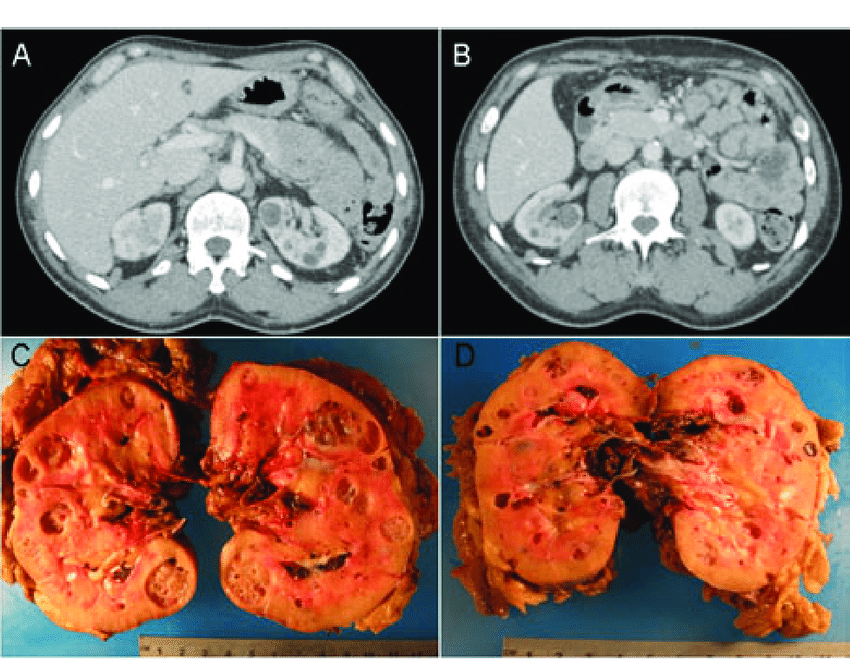
Acquired cystic kidney disease is a consequence of sustained uremia in patient with end-stage renal disease . The disease is found in 813% patients with end-stage renal disease and in approximately 50% patients on dialysis. The disease is multifactorial. It is the progressive destruction of renal functioning nephrons with compensatory hypertrophy of remaining renal parenchyma, obstruction of renal tubules by interstitial fibrosis or oxalate crystals, and cyst formation . Kidneys are atrophic and contain multiple cysts with variable size and imaging appearance . Since renal cysts are extremely common in the adult population, the diagnosis of ACKD requires the presence of three or more cysts in each kidney, in conjunction to end-stage renal disease, and no history of hereditable renal disease . Cyst hemorrhage is a common complication and can cause hematuria, whereas cyst rupture, perinephric hematoma, and retroperitoneal hemorrhage are less frequent . Development of RCC in the wall of the cyst is the most serious complication of ACKD, with a higher rate in comparison to the general population .
Fig. 12
Acquired cystic renal disease. Axial contrast-enhanced CT image shows atrophic kidneys, which contain multiple cysts of variable size
Multilocular Cystic Renal Neoplasm Of Low Malignant Potential

Last author update:Last staff update:Copyright:Page views in 2021:Page views in 2022 to date:Cite this page:
- “Neoplasm composed entirely of numerous cysts, the septa of which contain individual or groups of clear cells without expansile growth”
- Clear cells with low grade nuclei
- No recurrence or metastases when diagnosed on strict criteria
- Distinguish from regressing clear cell renal cell carcinoma with cystic degeneration, which often has cysts filled with hemorrhage, necrosis and hemosiderin deposits may have extensive hyalinization and often has areas of expansile growth of neoplastic cells
- 2016 WHO separates this neoplasm of low malignant potential from cystic renal cell carcinomas which have some overlapping morphologic features
- Use of “multilocular cystic renal cell carcinoma” is considered obsolete some literature references in this topic use the older terminology
- Accounts for less than 1% of all renal tumors
- Middle age adults
- Male:female ratio ranges from 1.2:1 to 2.1:1
- 90% are discovered incidentally, on radiology for other purposes
- Excellent prognosis with no recurrences or metastases in multiple case series when diagnosed on strict criteria
- There are no current guidelines or definitive evidence regarding the question of whether complete histologic examination is required for diagnosis
- Resection, preferably nephron sparing surgery
Large multi-cystic mass
Cysts filled with gelatinous material
Recommended Reading: Invasive Ductal Carcinoma Grade 3 Survival Rate
Localized Cystic Renal Disease
Localized cystic renal disease is a rare, nonhereditary, form of cystic renal disease, which manifests as a conglomeration of multiple simple cysts of variable size . In contrast to ACKD and ADPKD, localized cystic renal disease is typically unilateral and not progressive. The disease usually involves only a portion of the kidney with a polar predilection . Entire renal involvement is rare . The contralateral kidney is normal. The presence of interposed normal renal parenchyma and the absence of a capsule help to differentiate localized cystic renal disease from cystic nephroma and multiloculated cystic RCC . Cystic involvement of other organs is typically absent .
Fig. 13
Localized cystic renal disease. Axial contrast-enhanced CT image shows a conglomeration of multiple simple cysts of variable size in the right kidney
How Do Physicians Determine Staging Of Renal Cell Carcinoma
Staging of renal cell cancer is based on the size of the tumor and the extent of its spread outside the kidney. Like many cancers, renal cell cancer is staged according to the tumor , nodes , and metastases classification endorsed by the American Joint Committee on Cancer . The TNM classification system assigns a T code, an N code, and an M code to each tumor. An X is used if that feature cannot be determined. The combination of these 3 codes determines the disease stage.
- Primary tumor 0, 1, 2, 3a, 3b, 3c, 4 based on size of tumor and parts of kidney and surrounding area involved
- Regional lymph nodes 0, 1, 2 based on number of lymph nodes involved
- Distant metastasis 0 , 1
AJCC Stages
- Stage I T1N0M0 cancer is 7 cm or smaller in size and confined to the kidney
- Stage II T2N0M0 cancer is larger than 7 cm and confined to the kidney
- Stage III T1 or 2, N1M0 or T3a-c, N0-1, M0 cancer of any size that has spread to fatty tissue, blood vessels, or a lymph node near the kidney
- Stage IV T4 or any T, N2M0 or any T, any N, M1 cancer has spread to multiple lymph nodes to nearby organs, such as the bowel or pancreas or to other parts of the body, such as the lung, brain, or bone
Read Also: Amelanotic Melanoma Blanch
What Are The Types Of Kidney Cancer
The information in this document refers to renal cell carcinoma the most common form of kidney cancer. However, there are different types of kidney cancer, including:
- Renal cell carcinoma : This is the most common form of kidney cancer in adults and accounts for 85% of all kidney cancers. Renal cell carcinoma usually develops as a single tumor in one kidney, but it can affect both kidneys. Renal cell carcinoma begins in the cells that line the small tubes that are part of the nephrons within the kidneys. .
- Transitional cell carcinoma: Transitional cell carcinoma accounts for 6% to 7% of all kidney cancers. This cancer usually begins in the area where the ureter connects to the main part of the kidney. This area is called the renal pelvis. Transitional cell carcinoma also can occur in the ureters or bladder.
- Renal sarcoma: This is the least common form of kidney cancer, accounting for only 1% of kidney cancer cases. It begins in the connective tissues of the kidneys and, if not treated, can spread to nearby organs and bones.
- Wilms tumor: This is the most common type of kidney cancer in children. It accounts for about 5% of kidney cancers.
Adjuvant And Neoadjuvant Therapy
Adjuvant therapy, which refers to therapy given after a primary surgery, has not been found to be beneficial in renal cell cancer. Conversely, neoadjuvant therapy is administered before the intended primary or main treatment. In some cases neoadjuvant therapy has been shown to decrease the size and stage of the RCC to then allow it to be surgically removed. This is a new form of treatment and the effectiveness of this approach is still being assessed in clinical trials.
Dont Miss: Melanoma On Face Prognosis
Don’t Miss: Lobular Breast Cancer Stage 3
Other Types Of Kidney Cancers
Other types of kidney cancers include transitional cell carcinomas, Wilms tumors, and renal sarcomas.
Transitional cell carcinoma: Of every 100 cancers in the kidney, about 5 to 10 are transitional cell carcinomas , also known as urothelial carcinomas.
Transitional cell carcinomas dont start in the kidney itself, but in the lining of the renal pelvis . This lining is made up of cells called transitional cellsthat look like the cells that line the ureters and bladder. Cancers that develop from these cells look like other urothelial carcinomas, such as bladder cancer, when looked at closely in the lab. Like bladder cancer, these cancers are often linked to cigarette smoking and being exposed to certain cancer-causing chemicals in the workplace.
People with TCC often have the same signs and symptoms as people with renal cell cancer blood in the urine and, sometimes, back pain.
For more information about transitional cell carcinoma, see Bladder Cancer.
Wilms tumor : Wilms tumors almost always occur in children. This type of cancer is very rare among adults. To learn more about this type of cancer, see Wilms Tumor.
Renal sarcoma: Renal sarcomas are a rare type of kidney cancer that begin in the blood vessels or connective tissue of the kidney. They make up less than 1% of all kidney cancers.
Sarcomas are discussed in more detail in Sarcoma- Adult Soft Tissue Cancer.
Publication History And Summary Of Changes
1.5.1. Publication history
The EAU RCC Guidelines were first published in 2000. This 2022 RCC Guidelines document presents a substantial update of the 2021 publication.
1.5.2. Summary of changes
All chapters of the 2022 RCC Guidelines have been updated, based on the 2021 version of the Guidelines. References have been added throughout the document.
New data have been included in the following sections, resulting in changed evidence summaries and recommendations in:
5.4 Summary of evidence and recommendations for the diagnostic assessment of RCC
|
Recommendations |
You May Like: Grade 3 Cancer Treatment
What Is Cystic Renal Cell Carcinoma
4.9/5
Similarly, it is asked, can renal cysts become malignant?
A fluid-filled sac, called a cyst, is the most common growth found in a kidney. Cysts are mostly not cancerous. Solid kidney tumors can be benign, but most often are found to be cancer. Kidney cancer is one of the top 10 most common cancers diagnosed in the United States.
Furthermore, how treatable is renal cell carcinoma? Renal cell cancer, also called renal adenocarcinoma, or hypernephroma, can often be cured if it is diagnosed and treated when still localized to the kidney and to the immediately surrounding tissue. The probability of cure is directly related to the stage or degree of tumor dissemination.
Similarly one may ask, what is the life expectancy of someone with renal cell carcinoma?
Renal cell carcinoma life expectancy The American Cancer Society suggests that the prognosis is good for patients diagnosed with stage I or stage II RCC . The prognosis worsens as stage III and IV develop.
Does renal cell carcinoma spread?
Renal cell carcinoma can spread from a mass of cancer cells or tumor to other parts of your body. This process is called metastasis. Cancer cells spread into the tissue around the tumor in your kidney. The cancer moves from your kidney into your lymph system, which has vessels throughout the body.
Chemotherapy Immunologic Therapy Targeted Therapy

There are several medications approved for treatment of renal cell carcinoma:
- Chemotherapy destroys actively growing cells
- Immune therapy uses a process that triggers your immune system to destroy tumor cells
- Targeted therapy is a type of therapy that specifically destroys the tumor cells
All of these medications are powerful, and they may produce serious side effects during your treatment and recovery.
Read Also: Is Melanoma Bad
What Causes Renal Cell Carcinoma
Cancer occurs when abnormal cells begin to grow or divide uncontrollably. The abnormal cells crowd out normal cells, causing problems at the site where the cancer started. Cancer can sometimes spread to other organs as well. RCC beings in one or both kidneys.
The kidneys are bean-shaped organs that filter blood and create urine to dispose of the waste. Renal cell cancer affects the lining of small tubules in the kidneys, where the filtration takes place.
Most people have two kidneys one on each side of the lower spine, near the hips. However, its possible to live a healthy life with just one kidney. RCC usually involves one tumor in one kidney, but it also may involve multiple tumors in both kidneys.
Certain factors and lifestyle choices affect your risk of getting RCC. You may have an increased risk of RCC if you:
-
smoke
-
have high blood pressure
What Are Causes And Risk Factors Of Renal Cell Carcinoma
The exact cause of renal cell cancer has not been determined. A number of different factors seem to contribute to renal cell cancer. These risk factors include the following:
- Cigarettesmoking doubles the risk of renal cell cancer and contributes to as many as one-third of all cases. The more someone smokes, the greater the risk is of that person developing renal cell cancer.
- Obesity is a risk factor. As body weight increases, so does the risk of developing renal cell cancer. This is especially true in women.
- Occupational exposure to petroleum products, heavy metals, solvents, coke-oven emissions, or asbestos
- Cystic kidney disease associated with chronic renal insufficiency
- Cystic changes in the kidney and renal dialysis
- Tuberous sclerosis
In its early stages, renal cell cancer usually causes no noticeable symptoms. Symptoms may occur only when cancer grows and begins to press on surrounding tissues or spread to other parts of the body. The symptoms vary considerably from person to person. Some people never develop any symptoms before the disease is discovered the cancer is found when they undergo imaging tests, such as a CT scan, for another reason. In a study in the Journal of Urology, approximately 53% of people with localized renal cell carcinoma had no symptoms.
Signs and symptoms of renal cell cancer may include the following:
- Malaise
Dont Miss: Lobular Carcinoma Survival Rate
Also Check: Lobular Breast Cancer Survival Rates
How Is Renal Cell Carcinoma Diagnosed
If your doctor suspects you may have renal cell cancer, you will have a series of tests, including:
-
Physical exam: Your doctor will check for signs of disease, such as a lump near your kidney.
-
Health history: You will provide a detailed list of any conditions you and your family members have had. Your doctor will also ask about your health habits and lifestyle choices.
-
Imaging tests: Ultrasound, magnetic resonance imaging , and computed tomography scans take detailed pictures of your kidneys and other tissues inside the abdomen.
-
Blood tests: Your doctor may order tests that measure blood cell counts and kidney function.
-
Urinalysis: Urine tests check the urine for signs of protein and blood that could indicate cancer or other kidney diseases.
-
Biopsy: This definitive test for kidney cancer uses a thin needle to remove a bit of tissue. A pathologist examines the sample for evidence of cancer cells.
What Is Renal Cell Carcinoma
It’s the most common type of kidney cancer. Although itâs a serious disease, finding and treating it early makes it more likely that youâll be cured. No matter when youâre diagnosed, you can do certain things to ease your symptoms and feel better during your treatment.
Most people who have renal cell carcinoma are older, usually between ages 50 and 70. It often starts as just one tumor in a kidney, but sometimes it begins as several tumors, or itâs found in both kidneys at once. You might also hear it called renal cell cancer.
Doctors have different ways to treat renal cell carcinoma, and scientists are testing new ones, too. Youâll want to learn as much about your disease as you can and work with your doctor so you can choose the best treatment.
Read Also: Lobular Breast Cancer Survival Rate
What Is Clear Cell Renal Cell Carcinoma
Clear cell renal cell carcinoma, or ccRCC, is a type of kidney cancer. The kidneys are located on either side of the spine towards the lower back. The kidneys work by cleaning out waste products in the blood. Clear cell renal cell carcinoma is also called conventional renal cell carcinoma.
Clear cell renal cell carcinoma is named after how the tumor looks under the microscope. The cells in the tumor look clear, like bubbles.
Laparoscopic Nephrectomy And Robotic
These approaches to the operation are done through several small incisions instead of one large one. If a radical nephrectomy is needed, many doctors and patients now prefer to use these approaches when they can.
Laparoscopic nephrectomy: Special long instruments are inserted through the incisions, each of which is about 1/2-inch long, to remove the kidney. One of the instruments, the laparoscope, is a long tube with a small video camera on the end. This lets the surgeon see inside the abdomen. Usually, one of the incisions has to be made longer in order to remove the kidney .
Robotic-assisted laparoscopic nephrectomy: This approach uses a robotic system to do the laparoscopic surgery remotely. The surgeon sits at a panel near the operating table and controls robotic arms to operate. For the surgeon, the robotic system may allow them to move the instruments more easily and with more precision than during standard laparoscopic surgery. But the most important factor in the success of either type of laparoscopic surgery is the surgeons experience and skill. This is a difficult approach to learn. If you are considering this type of operation, be sure to find a surgeon with a lot of experience.
Don’t Miss: Stage 3 Cancer Symptoms