What Are Precancerous Cells
Precancerous cells are cells with an abnormal appearance suggestive of an increased cancer risk. These cells are not cancerous themselves, but can precede the development of cancer. When a patient has precancerous cells, they are an indicator that the patient should be monitored carefully in the future. Consistent screening and monitoring will help a doctor identify cancer early, if it shows up, allowing for prompt provision of treatment. Precancerous cells can also indicate the need for prophylactic treatment to prevent the appearance of cancer.
Such cells are identified in the laboratory by analyzing a sample of cells from the patient’s body. A doctor may take a cell biopsy if physical changes have been observed and there is a concern about cancer, or a biopsy may be taken as part of a routine medical screening like a Pap test for women. A lab technician will look at the cells under a microscope, examining them for signs of abnormalities.
The Five Stages Of Skin Cancer
Cancer in the skin thats at high risk for spreading shares features with basal cell carcinoma and squamous cell carcinoma. Some of these features are:
- Not less than 2 mm in thickness
- Has spread into the inner layers of the skin
- Has invaded skin nerves
Stage 0
In the earliest stage, cancer is only present in the upper layer of the skin. You may notice the appearance of blood vessels or a dent in the center of the skin growth. There are no traces of malignant cells beyond this layer.
Stage 1
At stage 1, cancer has not spread to muscles, bone, and other organs. It measures roughly 4/5 of an inch. Theres a possibility that it may have spread into the inner layer of the skin.
Stage 2
In this stage, cancer has become larger than 4/5 of an inch. Cancer still has not spread to muscles, bone, and other organs.
Stage 3
At stage 3, the cancer is still larger than 4/5 of an inch. Facial bones or a nearby lymph node may have been affected, but other organs remain safe. It may also spread to areas below the skin, such as into muscle, bone, and cartilage but not far from the original site.
Stage 4
Cancer can now be of any size and has likely spread into lymph nodes, bones, cartilage, muscle, or other organs. Distant organs such as the brain or lungs may also be affected. In rare cases, this stage might cause death when allowed to grow and become more invasive.
Are All Moles Cancerous
Most moles are not cancerous. Some moles are present at birth, others develop up to about age 40. Most adults have between 10 and 40 moles.
In rare cases, a mole can turn into melanoma. If you have more than 50 moles, you have an increased chance of developing melanoma.
A note from Cleveland Clinic
Your skin is the largest organ in your body. It needs as much attention as any other health concern. What may seem like an innocent cosmetic imperfection, may not be. Performing regular skin self-checks is important for everyone and is especially important if you are a person at increased risk of skin cancer. Skin cancer is also color-blind. If you are a person of color, skin cancer can happen to you. Check your skin every month for any changes in skin spots or any new skin growths. Consider taking skin selfies so you can easily see if spots change over time. If youre a person of color, be sure to check areas more prone to cancer development, such as the palms of your hands, soles of your feet, between your toes, your genital area and under your nails. Takes steps to protect your skin. Always wear sunscreen with SPF of at least 30 every day of the year. Wear UV-A/UV-B protective sunglasses, wide-brimmed hats and long-sleeve shirts and pants. See your dermatologist at least once a year for a professional skin check.
Last reviewed by a Cleveland Clinic medical professional on 11/19/2021.
References
Don’t Miss: What Is Large Cell Carcinoma
How Common Is Skin Cancer
Skin cancer is the most common cancer diagnosed in the U.S.
Other skin cancer facts:
- Around 20% of Americans develop skin cancer sometime in their life.
- Approximately 9,500 Americans are diagnosed with skin cancer every day.
- Having five or more sunburns in your life doubles your chance of developing melanoma. The good news is that the five-year survival rate is 99% if caught and treated early.
- Non-Hispanic white persons have almost a 30 times higher rate of skin cancer than non-Hispanic Black or Asian/Pacific Islander persons.
- Skin cancer in people with skin of color is often diagnosed in later stages when its more difficult to treat. Some 25% of melanoma cases in African Americans are diagnosed when cancer has spread to nearby lymph nodes.
Precancerous Types Of Skin Cancer

Some precancerous growths, often attributable to sun exposure, can lead to skin cancer over time. However, if they are recognized and removed early, you could avoid a cancer diagnosis. These growths include:
- Actinic keratosis: About 40-60% of squamous cell cancer cases began as actinic keratosis. Anywhere between 2-10% of these growths will develop into SCC, sometimes in as little as a couple of years. Actinic cheilitis is a type of actinic keratosis that appears on the lower lip, and is at higher risk for developing into skin cancer
- Bowens disease: This early, noninvasive form of SCC is at high risk of becoming skin cancer if not addressed. It presents as an eczema-like scaly patch and is usually red or brown in color. These growths have been linked to sun exposure, radiation, carcinogen exposure, genetics, and trauma
- Leukoplakia: These white patches on the lips, tongue, and gums may be caused by alcohol and tobacco use, and can turn into squamous cell carcinoma. Cancer sites on the lips may be caused by sun damage
- Keratoacanthoma: This dome-shaped growth is usually found on sun-exposed skin and usually grows quickly at first, then slows down. Many shrink and go away on their own, but if they continue to grow, this tumor can turn into squamous cell carcinoma. They are usually removed surgically
Don’t Miss: Carcinoma Causes
Rashes Linked To Other Cancers
A rash may also be a sign of cancers that develop away from the skin, such as different forms of lymphoma.
Lymphoma is dangerous, as cancer cells circulate throughout the body. These cells may then grow in many organs or tissues at once.
In the sections below, we list some other types of cancer that may cause skin symptoms:
How Is Actinic Keratosis Treated
Treatment for an actinic keratosis may include:
-
Cryotherapy. This treatment freezes the lesion.
-
Topical chemotherapy. This is medicine applied to the skin.
-
Laser surgery. This can remove lesions from the face and scalp, and actinic cheilitis from the lips.
-
Other treatments. These are done to remove or destroy the lesion.
Most actinic keratoses can be treated and cured. In rare cases they may come back. Its important to have regular skin exams after treatment. This will help check for new actinic keratoses and skin cancer.
You May Like: Merkel Cancer Prognosis
What Do Actinic Keratoses Look Like
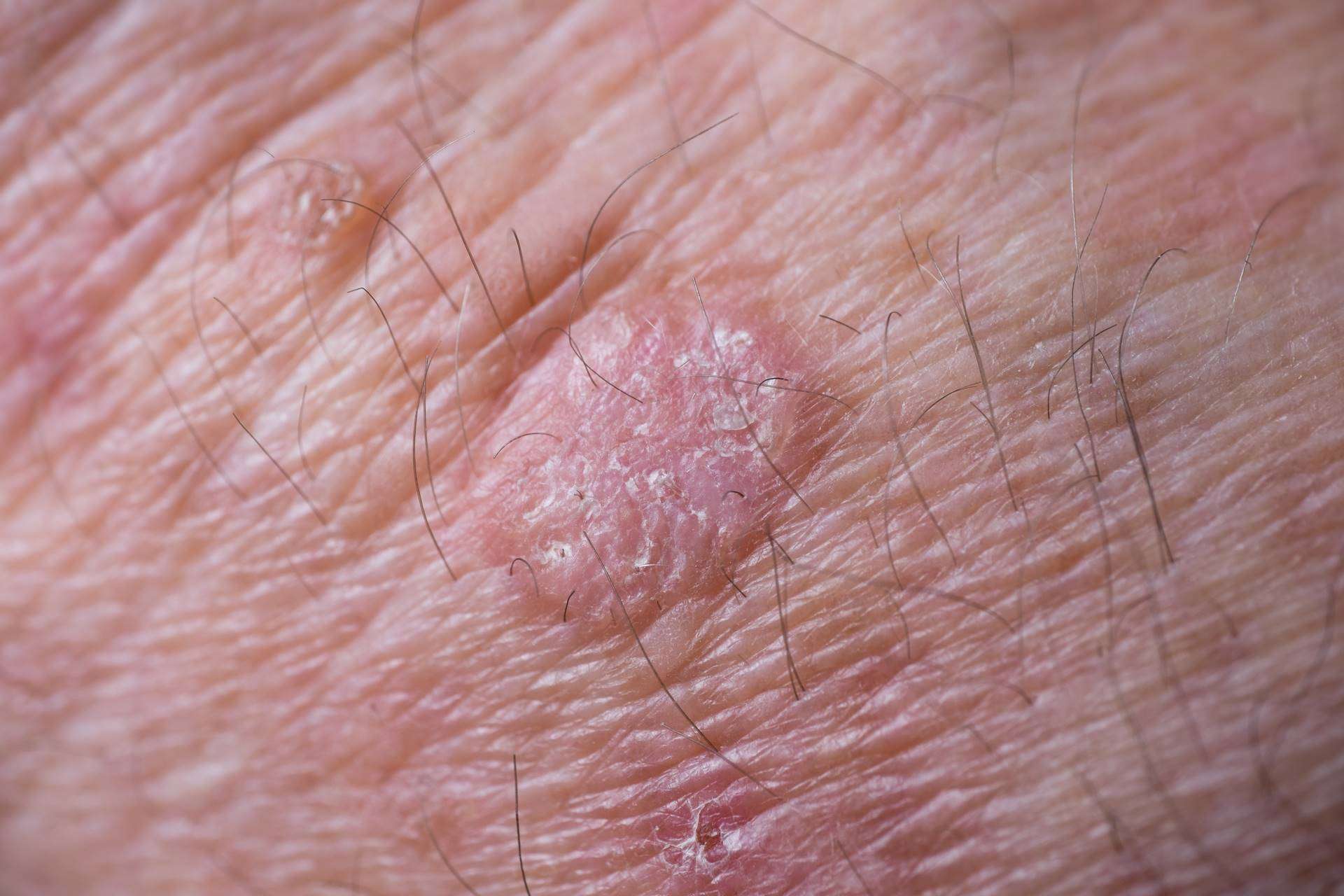
AKs often appear as small dry, scaly or crusty patches of skin. They may be red, light or dark tan, white, pink, flesh-toned or a combination of colors and are sometimes raised. Because of their rough texture, actinic keratoses are often easier to feel than see. For photos, go to our warning signs page.
What Is Skin Cancer
Skin cancer happens when skin cells grow and multiply in an uncontrolled, unorderly way.
Normally, new skin cells form when cells grow old and die or when they become damaged. When this process doesnt work as it should, a rapid growth of cells results. This collection of cells may be noncancerous , which dont spread or cause harm, or cancerous, which may spread to nearby tissue or other areas in your body if not caught early and treated.
Skin cancer is often caused by ultraviolet light exposure from the sun.
There are three main types of skin cancer:
Basal cell carcinoma and squamous cell carcinoma are the most common types of skin cancer and are sometimes called non-melanoma skin cancer.
Melanoma is not as common as basal cell or squamous cell carcinomas but is the most dangerous form of skin cancer. If left untreated or caught in a late-stage, melanomas are more likely to spread to organs beyond the skin, making them difficult to treat and potentially life-limiting.
Fortunately, if skin cancer is identified and treated early, most are cured. This is why it is important to take a few safeguards and to talk with your healthcare provider if you think you have any signs of skin cancer.
Don’t Miss: Clear Cell Carcinoma Symptoms
How Can I Help My Child Live With Skin Cancer
If your child has skin cancer, you can help him or her during treatment in these ways:
-
Your child may have trouble eating. A dietitian or nutritionist may be able to help.
-
Your child may be very tired. He or she will need to learn to balance rest and activity.
-
Get emotional support for your child. Counselors and support groups can help.
-
Keep all follow-up appointments.
-
Keep your child out of the sun.
After treatment, check your child’s skin every month or as often as advised.
When Is A Mole A Problem
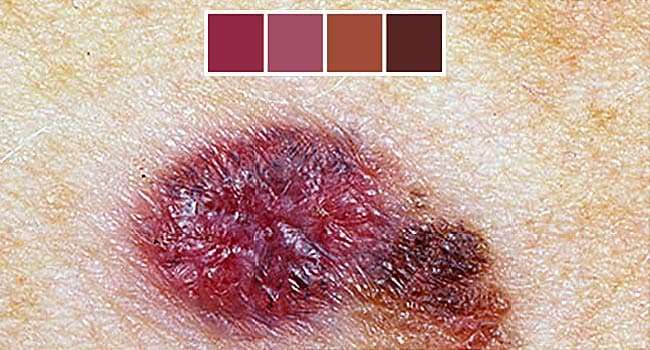
A mole is a benign growth of melanocytes, cells that gives skin its color. Although very few moles become cancer, abnormal or atypical moles can develop into melanoma over time. âNormalâ moles can appear flat or raised or may begin flat and become raised over time. The surface is typically smooth. Moles that may have changed into skin cancer are often irregularly shaped, contain many colors, and are larger than the size of a pencil eraser. Most moles develop in youth or young adulthood. Itâs unusual to acquire a mole in the adult years.
You May Like: How Serious Is Melanoma Skin Cancer
Don’t Miss: Malignant Breast Cancer Survival Rate
Skin Cancer Prevention In Portland
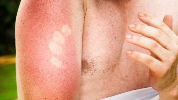
Most skin cancers result from too much sun exposure. The ultraviolet A rays can cause premature wrinkling, brown age spots while the ultraviolet B rays cause sunburns. Both types of radiation, UVA and UVB, are harmful and can cause skin cancer. It is essential to protect your skin from these damaging rays in order to prevent skin cancer. Not only do sunburns increase skin cancer, a tan is a sign of sun damage and will increase your risk of skin cancer.
What Causes Skin Cancer

The main cause of skin cancer is overexposure to sunlight, especially when it results in sunburn and blistering. Ultraviolet rays from the sun damage DNA in your skin, causing abnormal cells to form. These abnormal cells rapidly divide in a disorganized manner, forming a mass of cancer cells.
Another cause of skin cancer is frequent skin contact with certain chemicals, such as tar and coal.
Many other factors can increase your risk of developing skin cancer. See question, Who is most at risk for skin cancer?
Don’t Miss: Invasive Lobular Breast Cancer Survival Rate
What Are The Risk Factors For Actinic Keratosis
UV rays from the sun and from tanning beds cause almost all actinic keratoses. Damage to the skin from UV rays builds up over time. This means that even short-term exposure to sun on a regular basis can build up over a lifetime and increase the risk of actinic keratoses. Some people are more at risk than others, including:
-
People with pale skin, blonde or red hair, and blue, green, or gray eyes
-
People with darker skin, hair, and eyes who have been exposed to UV rays without protection
-
Older adults
-
People with suppressed immune systems
-
People with rare conditions that make the skin very sensitive to UV rays, such as albinism or xeroderma pigmentosum
Actinic Keratosis On An Arm
This photo contains content that some people may find graphic or disturbing.
Actinic keratosis, also called solar keratosis, is a precancerous skin lesion usually caused by too much sun exposure. It can also be caused by other factors such as radiation or arsenic exposure.
If left untreated, actinic keratoses can develop into a more invasive and potentially disfiguring skin cancer called squamous cell carcinoma. They appear predominantly on sun-exposed areas of the skin such as the face, neck, back of the hands and forearms, upper chest, and upper back. You can also develop keratoses along the rim of your ear.
Actinic keratosis is caused by cumulative skin damage from repeated exposure to ultraviolet light, including that found in sunshine. Over the years, the genetic material in your cells may become irreparably damaged and produce these pre-cancerous lesions. The lesions, like those seen here on the arm, can later become squamous cell carcinoma, a more invasive cancer.
You May Like: Stage 3b Melanoma Survival Rate
S Of Moles Nevus Actinic Keratosis Psoriasis
Angela Underwood’s extensive local, state, and federal healthcare and environmental news coverage includes 911 first-responder compensation policy to the Ciba-Geigy water contamination case in Toms River, NJ. Her additional health-related coverage includes death and dying, skin care, and autism spectrum disorder.
Not all skin blemishes are cancerous, nor will they all become cancerous in the future. If you are worried about a spot on your skin, this gallery of photographs can help you distinguish between cancerous, noncancerous, and precancerous lesions.
Of course, diagnosing skin cancer is far from straightforward, so if you have any doubts, contact your dermatologist or primary care physician as soon as possible.
What Are The Symptoms Of Actinic Keratosis
Actinic keratosis develops slowly. It most likely appears on areas of skin often exposed to the sun. These can include the face, ears, bald scalp, neck, backs of hands and forearms, and lips. It tends to lie flat against the skin of the head and neck, but appears as a bump on arms and hands. The base of an actinic keratosis may be light or dark, tan, pink, red, or a combination of these. Or it may be the same color as the skin. The scale or crust may be horny, dry, and rough. In some cases, it may itch or have a prickly or sore feeling.
Often, a person will have more than one actinic keratosis lesion. Actinic keratoses that develop on the lip are called actinic cheilitis.
Don’t Miss: Melanoma Stage 3 Symptoms
Invasive Squamous Cell Cancer Of The Vulva
Almost all women with invasive vulvar cancers will have symptoms. These can include:
- An area on the vulva that looks different from normal it could be lighter or darker than the normal skin around it, or look red or pink.
- A bump or lump, which could be red, pink, or white and could have a wart-like or raw surface or feel rough or thick
- Thickening of the skin of the vulva
- Itching
- Bleeding or discharge not related to the normal menstrual period
- An open sore
Verrucous carcinoma, a subtype of invasive squamous cell vulvar cancer, looks like cauliflower-like growths similar to genital warts.
These symptoms are more often caused by other, non-cancerous conditions. Still, if you have these symptoms, you should have them checked by a doctor or nurse.
What Is The Difference In Precancerous Skin Growths And Skin Cancer
Categories:Cancer Prevention,Skin Cancer
May 20, 2021
Have you ever wondered what causes a common mole to develop into skin cancer? Most moles never cause problems and don’t progress to skin cancer.
There are a number of reasons you may want to have a doctor or dermatologist look at a skin growth:
- Its new to you
- Its a former mole thats growing or spreading
- The growth is irritated or hurts
- There is bleeding from the growth
- It looks like a sore that won’t heal
Because you have a new spot or there has been a change doesnt automatically mean its cancerous. Lets talk about what you should be looking for and what to do if you notice something different on your skin.
Recommended Reading: Well Differentiated Squamous Cell Carcinoma Stages
The Early Stages Of Skin Cancer
Some forms of cancer, especially melanoma, may appear suddenly and without warning. Most people become alarmed only when they develop a crust or sore that refuses to heal. Did you know that the early stages of cancer do not always look or feel so bad? Harmless-looking moles, skin lesions, or unusual skin growths may also be the signs of early stages.
Regular skin examination can help you spot these early clues. If you see anything suspicious or observe unusual appearances in your skin, we can help you get the right diagnosis and treatment immediately. Some forms of cancer in the skin can be life-threatening and spread without being given urgent attention.
Tests Or Procedures That Examine The Skin Are Used To Diagnose Basal Cell Carcinoma And Squamous Cell Carcinoma Of The Skin

The following procedures may be used:
- Physical exam and health history: An exam of the body to check general signs of health, including checking for signs of disease, such as lumps or anything else that seems unusual. A history of the patients health habits and past illnesses and treatments will also be taken.
- Skin exam: An exam of the skin for bumps or spots that look abnormal in color, size, shape, or texture.
- Skin biopsy: All or part of the abnormal-looking growth is cut from the skin and viewed under a microscope by a pathologist to check for signs of cancer. There are four main types of skin biopsies:
- Shave biopsy: A sterile razor blade is used to shave-off the abnormal-looking growth.
- Punch biopsy: A special instrument called a punch or a trephine is used to remove a circle of tissue from the abnormal-looking growth. Enlarge Punch biopsy. A hollow, circular scalpel is used to cut into a lesion on the skin. The instrument is turned clockwise and counterclockwise to cut down about 4 millimeters to the layer of fatty tissue below the dermis. A small sample of tissue is removed to be checked under a microscope. Skin thickness is different on different parts of the body.
- Incisional biopsy: A scalpel is used to remove part of a growth.
- Excisional biopsy: A scalpel is used to remove the entire growth.
Recommended Reading: Cancer All Over Body Symptoms