Primary Vs Secondary Skin Lesions
Skin lesions are either primary or secondary. Primary skin lesions are either present from birth or develop during your lifetime.
Secondary skin lesions arise from primary skin lesions. This can happen when a primary skin lesion is:
- Disturbed
- Irritated
- Changes over time
For example, if eczema is scratched, a crust may form. The crust is a secondary lesion.
Image Gallery: Primary Skin Lesions
Alexander Werner Resnick, VMD, DACVD, Animal Dermatology Center
Alexander Werner Resnick, VMD, DACVD, is the editor of the dermatology section of the 5-Minute Veterinary Consult textbook and coauthor of the second and third editions of Small Animal Dermatology Clinical Companion. He practices at the Animal Dermatology Center in Studio City and Westlake Village, California, and Reno, Nevada.
This is a filled error message
To access full articles on www.cliniciansbrief.com, please sign in below.
For free.
Create an account for free
Want free access to the #1 publication for diagnostic and treatment information? Create a free account to read full articles and access web-exclusive content on www.cliniciansbrief.com.
The diagnosis of every disorder, including skin-related conditions, requires a systematic approach to a differential diagnosis list. For dermatologic diseases, abnormalities are visually apparent thus, an understanding of basic skin lesions and their patterns of presentation can improve the differential diagnosis list and diagnostic investigation plan.
Dermatologic diseases are often chronic or recurrent. With contemporary electronic medical records, concise and correct documentation of lesions allows for more accurate recording of disease progression and/or patient response to treatment.
Figure 7Nodule. A nodule is a deep, solid accumulation of cells in the skin. Nodules are typically larger than 1 cm.
What Are The Malignant Lesions Of Skin
Malignant lesions of the skin are common. Patients who develop squamous cell carcinoma and malignant melanoma often have recognizable precursor conditions. A few skin lesions resemble malignancies. Lesions that are growing, spreading or pigmented, or those that occur on exposed areas of skin are of particular concern.
Also Check: Which Is Worse Basal Or Squamous Cell Skin Cancer
What Is A Benign Skin Lesion
A benign skin lesion is a non-cancerous skin abnormality, , or tumor that can occur anywhere on the . Benign lesions can manifest in a number of different ways, depending on their cause and tissue of origin. Common benign skin lesions include most , better known as , , skin tags, cherry angiomas, and lipomas, among others. Most of the time, these lesions are harmless and dont require treatment, unless they cause symptoms such as discomfort or itching.
What Are The Primary Lesions Of The Skin

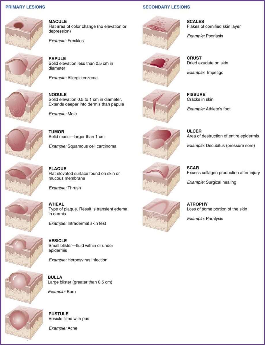
Primary lesions, which are associated with specific causes on previously unaltered skin, occur as initial reactions to the internal or external environment. Vesicles, bullae, and pustules are formed by fluid within skin layers. Nodules, tumors, papules, wheals, and plaques are palpable, elevated, solid masses.
Also Check: Can You Get Skin Cancer On Your Breast
What Are The Most Important Facts To Know About Skin Lesions
Skin lesions refer to any skin area that presents different characteristicsincluding , shape, size, and texturefrom the surrounding skin. Skin lesions can be hereditary, such as or birthmarks, or acquired as a result of , medications, sun exposure, and systemic diseases, such as autoimmune diseases, some infectious diseases, and cancer, among others. Diagnosis of skin lesions begins with physical examination and medical history, and some skin lesions may require further diagnostic tests, such as blood tests, imaging, or biopsy. Specific treatment depends on the type of lesion and if malignancy is present. Some benign lesions may not need to be treated at all, while others may need local treatment. If the skin lesion is caused by a systemic disease, treatment may also address the underlying cause. On the other hand, malignant and lesions are generally treated with surgical removal to prevent their progression. Finally, the use of protective sunscreen is recommended for all individuals.
First Of All What Is A Skin Lesion
A skin lesion is a broad term that refers to any abnormality on your skin. Medical dictionaries define skin lesion as a superficial growth or patch of the skin that does not resemble the area surrounding it. A skin lesion can be a rash, mole, wart, cyst, blister, bump, discoloration, or other change that you may notice on your skin.
A skin lesion can be a result of a simple scrape or cut or as severe as a pre-cancerous mole or mark. While the spectrum of lesions ranges significantly, there are some general categories you can use to identify yours.
Recommended Reading: How Can You Get Skin Cancer
So What Causes Skin Lesions
What causes skin lesions can range widely, depending on the specific type of lesion you have. The most common cause of skin lesions is an infection of the skin by bacteria, viruses, fungi or parasites transferred by touch or through the air. Warts and chickenpox are some examples of possible infections. Other types of lesions can be caused by allergic sensitivities or health conditions, such as diabetes or poor circulation. Skin lesions can also be a result of genetic predispositions . For example, some people are more likely to develop moles and freckles than others.
You should consult your doctor if you notice any suspicious skin lesions on your skin. A doctor will examine the lesion, looking for distinguishing characteristics and checking your past medical history. Doctors often also take a scrape or swab of the lesion to investigate the potential cause of infection.
Can Cancer Cause Skin Lesions
Cancer can cause skin lesions through the spreading of malignant cells to the skin or, more commonly, as a result of paraneoplastic syndromes, which are distant clinical manifestations triggered by an internal malignancy. Examples of cutaneous paraneoplastic syndromes include and . Other paraneoplastic conditions that involve the skin include Sweets syndrome, which causes skin lesions along with sudden onset fever, and the LeserTrélat sign, which includes the presence of multiple .
Read Also: How To Spot Melanoma Cancer
Principles Of Cutaneous Diagnosis
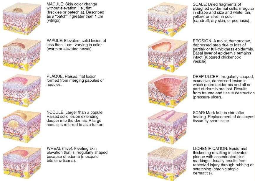
At the initial physical examination, all cutaneous abnormalities should be noted, using only appropriate morphologic descriptive terms. Any skin lesion can be described by one of the following generic nouns: macule, papule, plaque, nodule, tumor, vesicle, bulla, pustule, wheal, telangiectasis, comedo, burrow, or cyst. These are the primary lesions: the morphologic changes most representative of the pathologic process and the basis of the diagnostic categories of dermatologic disease. Primary lesions are discovered by diligent examination, not by history they do not represent the first lesions experienced by the patient. When primary lesions have been altered by external factors, secondary changes are seen: scale, crust, fissure, erosion, ulcer, excoriation, atrophy, scar. Appropriate adjectives such as color , surface , and feel can be added to the primary and secondary terms to evoke an accurate image in the minds of those reading the database. Note that an “accurate image” does not imply a diagnosis, only generic descriptive terminology.
When examining patients without overt dermatologic problems, the examiner should always note skin, hair, and nail abnormalities, as well as lesions such as scars, pigmentation, or moles that may be peripheral to the chief complaint but important to the patient’s medical evaluation.
Skin Lesions: What Do They Mean
While some people claim that all skin diseases look alike, the fact is to the trained eye, even subtle difference in skin changes can offer clues to the underlying disease process. One of the first steps in appreciating and understanding the differences of skin lesions is to learn what primary and secondary skin lesions actually are, and what they represent.
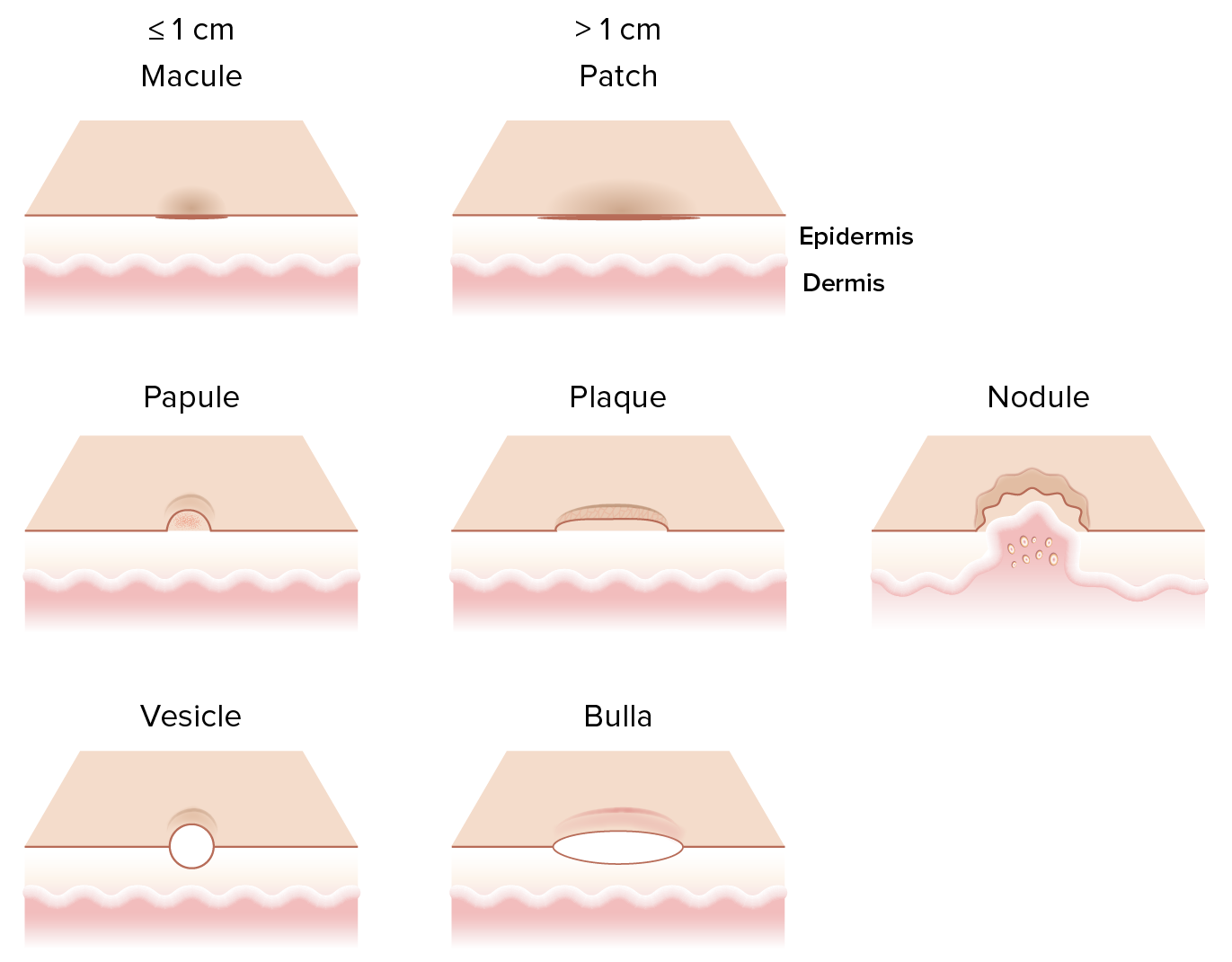
Macules are color changes of the skin less in one centimeter in diameter that do not have any substance or mass. Patches are larger than a centimeter. Most macules and patches are inflamed but may be hyper or hypopigmented.
Papules are solid, usually firm raised areas of the skin smaller than a centimeter. They are usually the result of a local irritation to the skin. Papules will develop due to skin infections , parasites , environmental or contact allergy. Sometimes papules are seen which then develop into a pustule. Papules are usually scattered and random within an inflamed area on the skin, but sometimes they are grouped into patterns such as circles.
Lichenification which is incorrectly referred to as elephant skin represents skin which has thickened as a result of chronic trauma, usually scratching. Lichenified skin can frequently be secondarily infected with Malassezia or Staphylococcus, which exacerbates the pruritus and leads to even more scratching and lichenification. Lichenified skin frequently is also hyperpigmented.
Related Content:
Recommended Reading: What Color Ribbon Is For Melanoma Cancer
Types Of Secondary Skin Lesions
Secondary skin lesions, which get inflamed and irritated, develop after primary skin lesions or due to an injury. The most common secondary skin lesions include
- Crust: A crust or a scab is a type of skin lesion that forms over a scratched, injured or irritated primary skin lesion. It is formed from the dried secretions over the skin.
- Ulcer: Ulcers are a break in the continuity of the skin or mucosa. Skin ulcers are caused by an infection or trauma. Poor blood circulation, diabetes, smoking and/or bedridden status increase the risk of ulcers.
- Scales: Scales are patches of skin cells that build up and flake off the skin. Patches are often seen in psoriasis and cause bleeding when they are removed.
- Scar: Injuries, such as scratches, cuts and scrapes, can leave scars. Some scars may be thick and raised. These may cause itching or oozing and appear reddish or brownish. These are called keloids.
- Skin atrophy: Skin atrophy occurs when areas of the skin become thin and wrinkled. This could occur due to the frequent use of steroid creams, radiation therapy or poor blood circulation.
How Many Types Of Skin Lesions Do Exist

In a nutshell, there are as many as two types of skin lesions named primary and secondary. You might be wondering the difference between the two.
- Primary Skin Lesions
- Secondary Skin Lesions
Primary skin lesions are those that are marked by the changes either in their texture or colour that a person may acquire over time. They can also be marked at the time of birth.
Primary skin lesions basically include an age spot or a birthmark .
On the reverse side of this, secondary skin lesions happen to be the progression of primary skin lesions.
Most of the times, they happen to be the changes to the original one resulting from the natural evolution.
Recommended Reading: What Is The Treatment For Melanoma
Skin Lesions & Tumors
A skin lesion is a part of the skin that has an abnormal growth or appearance compared to the skin around it. There are two categories of skin lesions, primary and secondary. Primary skin lesions are present at birth or are acquired over your lifetime. A birthmark would be an example of a primary skin lesion. Secondary skin lesions evolve from primary lesions or develop as a consequence of your activities. Melanoma resulting from sun exposure would be an example of a secondary skin lesion.
At Great Lakes ENT Specialists, we diagnose and treat all types of facial skin lesions with an emphasis on facial skin cancer.
What Is A Primary Skin Lesion
Skin lesions can be divided into two main types: primary and secondary. Primary skin lesions originate on previously healthy skin and are directly associated with a specific cause. Common examples of primary skin lesions include , , and blisters, among others. On the other hand, secondary skin lesions develop from the of a primary skin lesion, either due to traumatic manipulation, such as scratching or rubbing, or due to its treatment or progression. Examples of secondary skin lesions include crusts, sores, , and scars.
Join millions of students and clinicians who learn by Osmosis!
Don’t Miss: What Can Melanoma Look Like
When To See A Doctor
If OTC products do not resolve acne, eczema, or psoriasis, a person should contact a doctor, who may prescribe medication in the forms of creams, lotions, or pills.
There are no OTC treatments for impetigo. Anyone who thinks that they or their child has the infection should speak to a doctor.
Ringworm on the scalp requires medical attention. Anyone who suspects that they have this should see a doctor, who can prescribe antifungal medication.
Anyone who notices new moles or changes in existing moles should contact a doctor, who may screen for skin cancer. The same is true for people who have actinic keratosis.
What Is A Malignant Skin Lesion
A malignant skin lesion is, by definition, . The two main types of skin cancer are keratinocyte carcinoma and . Each type of skin cancer has unique characteristics, but general signs of skin cancer can include rapidly growing skin lesions, changes in the or size of a preexisting lesion, or a scabbing sore that doesnt heal with time.
You May Like: How To Know If You Have Melanoma
Definitions Of Primary And Secondary Lesions
Primary skin lesions are those which develop as a direct result of the disease process. Secondary lesions are those which evolve from primary lesions or develop as a consequence of the patient’s activities.
This classification is naturally artificial the same lesion type might be a primary lesion in one disease but a secondary lesion in another .
Do not confuse the term “secondary lesion” with “secondary pyoderma”. The latter term implies a bacterial infection which is complicating an underlying skin disease but that secondary pyoderma may present with primary lesions such as papules and pustules.
Examples Of Benign And Potentially Cancerous Lesions
Casey Gallagher, MD, is board-certified in dermatology. He is a clinical professor at the University of Colorado in Denver, and co-founder and practicing dermatologist at the Boulder Valley Center for Dermatology in Colorado.
Skin lesions are abnormal changes of the skin compared to the surrounding tissue. Skin lesions may look like bumps or patches, or they may be smooth. They may be a different color or texture compared to nearby skin.
There are many different types of skin lesions that you can be born with or acquire. Some are benign, which means they are harmless. Others can be severe and cancerous. They may appear all over your body, or they may be in one place.
The shape can vary, too. Some lesions are symmetrical, meaning they are the same shape all the way around. Others are irregular in shape.
The way a skin lesion looks and where it appears can help identify it. To find the cause of a skin lesion, healthcare providers consider:
- Color
This article looks at 20 different types of skin lesions, their causes, and their treatment.
Read Also: How To Look For Melanoma
Ways To Diagnose A Skin Lesion
A dermatologist makes a diagnosis of a skin lesion after a thorough physical examination and detailed medical history.
If examining your skin doesn’t provide clear results, your doctor may conduct tests, such as:
Dermatitis Due To Contact

- Itchy, red, scaly, or raw skin is a symptom of contact dermatitis.
- It manifests itself hours to days after coming into touch with an allergen.
- A contact dermatitis rash has distinct borders and occurs where your skin came into contact with the irritant.
- It also causes blisters to leak, ooze, or crust over.
Don’t Miss: Is Melanoma Caused By Sun Exposure
Chapter 103an Overview Of The Skin And Appendages
Dermatologic diagnosis depends so much on accurate and complete description of cutaneous findings that an inexperienced but observant medical student who is careful and complete may discover a critical clue overlooked by a competent but hurried examining physician. In evaluating cutaneous disorders, objective findings from the physician’s physical examination and office diagnostic tests are weighted more heavily than the patient’s subjective history. An initial brief patientphysician interchange establishes rapport and defines the complaint, but a greater proportion of time should be spent on a Sherlockian physical examination of the entire integument of a new patient.
What Causes Lesions On The Skin
- An infection on or in the skin is the most prevalent cause of a skin lesion.
- A wart is one example. The human papillomavirus , which causes warts, is transmitted from person to person via direct skin-to-skin contact. Direct touch is also used to spread the herpes simplex virus, which causes both cold sores and genital herpes.
- Skin lesions can appear all over your body as a result of a systemic infection, which is an infection that spreads throughout your body. Chickenpox and shingles are two examples. MRSA and cellulitis are two potentially fatal illnesses that cause skin sores.
- Moles and freckles, for example, are skin blemishes that are inherited. Birthmarks are lesions that are present at birth.
- Others, such as allergic eczema and contact dermatitis, can be the result of an allergic reaction. Some medical disorders, such as poor circulation or diabetes, create skin sensitivity, which can result in lesions.
Recommended Reading: What Is Invasive High Grade Urothelial Carcinoma
How Do You Diagnose Skin Lesions
Diagnosis of skin lesions begins with careful physical examination and medical history. Physical examination involves assessing the , size, shape, depth, location, and comparison with other lesions. can be performed to examine skin lesions under a magnifying glass. A Woods lamp examination can also be used to evaluate certain skin conditions under a black light. Additionally, certain aspects of the medical history can offer valuable information to guide the diagnosis, including sun exposure, , , contact with irritants, previous malignancy, and family history.
Some skin lesions may require further diagnostic tests. These can include blood tests, tests, skin or wound swabs for microbiological investigations, and imaging techniques, such as an X-ray or CT scan. Finally, if the diagnosis is still uncertain or malignancy is suspected, a biopsy can be performed.