Where Does Skin Cancer Develop
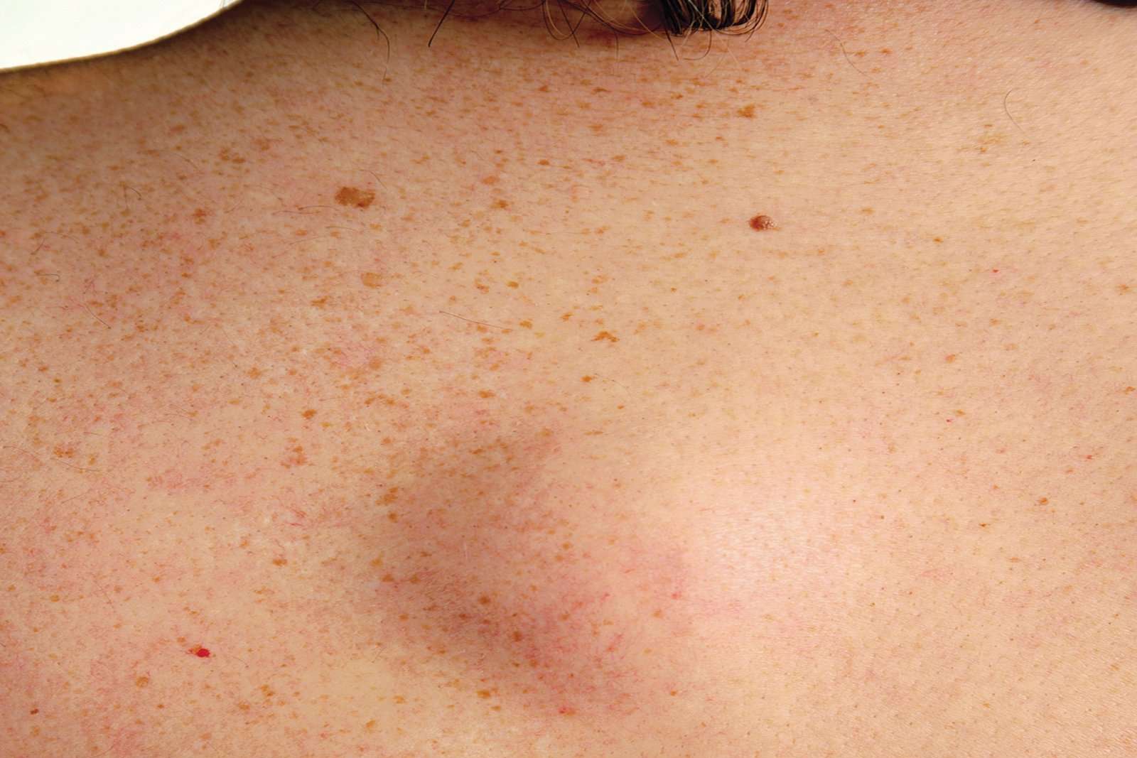
Skin cancer is most commonly seen in sun-exposed areas of your skin your face , ears, neck, arms, chest, upper back, hands and legs. However, it can also develop in less sun-exposed and more hidden areas of skin, including between your toes, under your fingernails, on the palms of your hands, soles of your feet and in your genital area.
What Are Skin Cancers Of The Feet
Skin cancer can develop anywhere on the body, including in the lower extremities. Skin cancers of the feet have several features in common. Most are painless, and often there is a history of recurrent cracking, bleeding, or ulceration. Frequently, individuals discover their skin cancer after unrelated ailments near the affected site.
What Causes Skin Cancer
The main cause of skin cancer is overexposure to sunlight, especially when it results in sunburn and blistering. Ultraviolet rays from the sun damage DNA in your skin, causing abnormal cells to form. These abnormal cells rapidly divide in a disorganized manner, forming a mass of cancer cells.
Another cause of skin cancer is frequent skin contact with certain chemicals, such as tar and coal.
Many other factors can increase your risk of developing skin cancer. See question, Who is most at risk for skin cancer?
You May Like: What Is The Survival Rate For Invasive Lobular Carcinoma
Curettage Electrodesiccation And Cryotherapy
Some dermatologists perform curettage, electrodesiccation, and cryotherapy to treat skin cancer. These are considered to be destructive techniques that are best suited for small, superficial carcinomas with definite borders. During the procedure, layers of skin cells are scraped away using a curette. Any remaining cancer cells are destroyed with the use of an electric needle.
In some cases, liquid nitrogen or cryotherapy is used to freeze the margins of the treatment area. Extremely low temperatures kill the malignant skin cells and create a wound, which will heal in a few weeks. The treatment may leave scars that are flat and round, similar to the size of the skin cancer lesion.
Skin Cancer: Prevention Treatment And Signs Of Melanoma

17 October 14
One in five Americans develops skin cancer over their lifetime, making it the most common form of cancer. Fortunately it is also one of the most preventable, because sun exposure is a major factor in its growth, according to the American Academy of Dermatology .
âPeople of every skin color can expect that they will be at risk of developing skin cancer,â said Dr. Doris Day, a board-certified dermatologist New York City and an attending physician at Lenox Hill Hospital, also in New York. âBut the good news is, that if caught early, greater than 98 percent of skin cancers are curable, and sometimes not even with surgery.â
Donât Miss: Invasive Ductal Carcinoma Grade 3 Life Expectancy
You May Like: Lobular Carcinoma Survival Rate
What Causes Skin Cancer In A Child
Exposure to sunlight is the main factor for skin cancer. Skin cancer is more common in people with light skin, light-colored eyes, and blond or red hair. Other risk factors include:
-
Age. Your risk goes up as you get older.
-
Family history of skin cancer
-
Having skin cancer in the past
-
Time spent in the sun
-
Using tanning beds or lamps
-
History of sunburns
-
Having atypical moles . These large, oddly shaped moles run in families.
-
Radiation therapy in the past
-
Taking a medicine that suppresses the immune system
-
Certain rare, inherited conditions such as basal cell nevus syndrome or xeroderma pigmentosum
-
HPV infection
-
Actinic keratoses or Bowen disease. These are rough or scaly red or brown patches on the skin.
Squamous Cell Carcinoma Early Stages
The second most common form of cancer in the skin is squamous cell carcinoma. At first, cancer cells appear as flat patches in the skin, often with a rough, scaly, reddish, or brown surface. These abnormal cells slowly grow in sun-exposed areas. Without proper treatment, squamous cell carcinoma can become life-threatening once it has spread and damaged healthy tissue and organs.
Also Check: Invasive Ductal Carcinoma Survival Rate Stage 1
Basal Cell And Squamous Cell Carcinomasigns And Symptoms
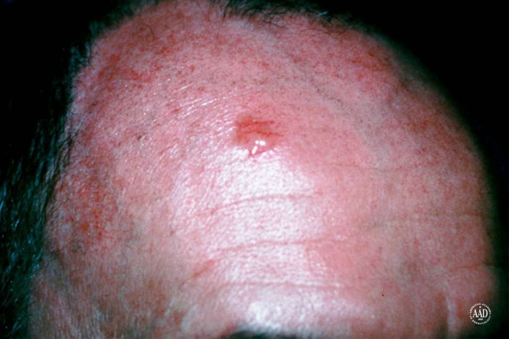
The most common warning sign of skin cancer is a change on the skin, especially a new growth or a sore that doesn’t heal. The cancer may start as a small, smooth, shiny, pale or waxy lump. It also may appear as a firm red lump. Sometimes, the lump bleeds or develops a crust.
Both basal and squamous cell cancers are found mainly on areas of the skin that are exposed to the sun the head, face, neck, hands and arms. But skin cancer can occur anywhere.
An early warning sign of skin cancer is the development of an actinic keratosis, a precancerous skin lesion caused by chronic sun exposure. These lesions are typically pink or red in color and rough or scaly to the touch. They occur on sun-exposed areas of the skin such as the face, scalp, ears, backs of hands or forearms.
Actinic keratoses may start as small, red, flat spots but grow larger and become scaly or thick, if untreated. Sometimes they’re easier to feel than to see. There may be multiple lesions next to each other.
Early treatment of actinic keratoses may prevent them from developing into cancer. These precancerous lesions affect more than 10 million Americans. People with one actinic keratosis usually develop more. Up to 1 percent of these lesions can develop into a squamous cell cancer.
Basal cell carcinoma is the most commonly diagnosed skin cancer. In recent years, there has been an upturn in the diagnoses among young women and the rise is blamed on sunbathing and tanning salons.
- Raised, dull-red skin lesion
Basal Cell Carcinoma Early Stages
Basal cells are found within the skin and are responsible for producing new skin cells as old ones degenerate. Basal cell carcinoma starts with the appearance of slightly transparent bumps, but they may also show through other symptoms.
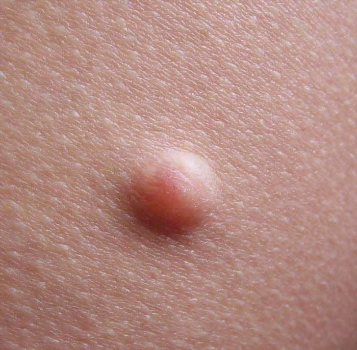
In the beginning, a basal cell carcinoma resembles a small bump, similar to a flesh-colored mole or a pimple. The abnormal growths can also look dark, shiny pink, or scaly red in some cases.
Also Check: Web Md Skin Cancers
More Pictures Of Basal Cell Carcinoma
While the above pictures show you some common ways that BCC can appear on the skin, this skin cancer can show up in other ways, as the following pictures illustrate.
Scaly patch with a spot of normal-looking skin in the center
On the trunk, BCC may look like a scaly patch with a spot of normal-looking skin in the center and a slightly raised border, as shown here.
Basal cell carcinoma can be lighter in some areas and darker in others
While BCC tends to be one color, it can be lighter in some areas and darker in others, as shown here.
Basal cell carcinoma can be brown in color
Most BCCs are red or pink however, this skin cancer can be brown, as shown here.
Basal cell carcinoma can look like a group of shiny bumps
BCC can look like a group of small, shiny bumps that feel smooth to the touch.
Basal cell carcinoma can look like a wart or a sore
The BCC on this patients lower eyelid looks like a wart* in one area and a sore** in another area.
If you see a spot or growth on your skin that looks like any of the above or one that is growing or changing in any way, see a board-certified dermatologist.
When To Visit A Podiatrist
Podiatrists are uniquely trained as lower extremity specialists to recognize and treat abnormal conditions on the skin of the lower legs and feet. Skin cancers affecting the feet may have a very different appearance from those arising on the rest of the body. For this reason, a podiatrist’s knowledge and clinical training is of extreme importance for patients for the early detection of both benign and malignant skin tumors.
Learn the ABCDs of melanoma. If you notice a mole, bump, or patch on the skin that meets any of the following criteria, see a podiatrist immediately:
- Asymmetry – If the lesion is divided in half, the sides don’t match.
- Borders – Borders look scalloped, uneven, or ragged.
- Color – There may be more than one color. These colors may have an uneven distribution.
- Diameter The lesion is wider than a pencil eraser .
To detect other types of skin cancer, look for spontaneous ulcers and non-healing sores, bumps that crack or bleed, nodules with rolled or donut-shaped edges, or scaly areas.
Read Also: Stage 2 Invasive Ductal Carcinoma Survival Rate
The Four Major Types Of Melanoma
Melanoma can be divided into different subtypes. A few of the most common subtypes are:
- Superficial spreading melanoma.Superficial spreading melanoma is the most common type of melanoma. Lesions are usually flat, irregular in shape, and contain varying shades of black and brown. It can occur at any age.
- Lentigo maligna melanoma. Lentigo maligna melanoma usually affects adults over 65 and involves large, flat, brownish lesions.
- Nodular melanoma.Nodular melanoma can be dark blue, black, or reddish-blue, but may have no color at all. It usually starts as a raised patch.
- Acral lentiginous melanoma.Acral lentiginous melanoma is the least common type. Typically it affects the palms, soles of the feet, or under finger and toenails.
What Are The Symptoms Of Skin Cancer Of The Head And Neck

Skin cancers usually present as an abnormal growth on the skin. The growth may have the appearance of a wart, crusty spot, ulcer, mole or sore. It may or may not bleed and can be painful. If you have a preexisting mole, any change in the characteristics of this spot – such as a raised or an irregular border, irregular shape, change in color, increase in size, itching or bleeding – are warning signs of melanoma. Sometimes the first sign of melanoma or squamous cell cancer is an enlarged lymph node.
Johns Hopkins Head and Neck Cancer Surgery Specialists
Our head and neck surgeons and speech language pathologists take a proactive approach to cancer treatment. Meet the Johns Hopkins specialists who will work closely with you during your journey.
Read Also: Invasive Ductal Carcinoma Prognosis
When To Talk To A Doctor About Your Lumps
Diagnosing lumps and bumps on your own can be challenging. If you are worried about cancer or have a history of cancer in your family, talk to us about it and we will answer your question: when to worry about a lump under the skin?
Cancer or other serious lumps will have these signs:
- Firm/hard to the touch
- It doesnt move around, fixed to the tissue
- Not tender when touched
- Felt in the breast or groin region
- Grows steadily
- Uneven surface
- New lump
One of my patients, Calla, agreed to share her experience when she presented with a similar complaint.
A Sore That Doesnt Heal
Many skin cancers are first dismissed as being due to a bug bite, minor injury, or irritation, but become more obvious when they dont go away over time. If you notice a sore on your skin that refuses to heal, even if it seems to be healing but then reappears, talk to your healthcare provider. In general, any skin change that hasnt resolved on its own over a period of two weeks should be evaluated.
Don’t Miss: Stage 3 Melanoma Survival Rate
Abcde Melanoma Detection Guide
A is for Asymmetry
Look for spots that lack symmetry. That is, if a line was drawn through the middle, the two sides would not match up.
B is for Border
A spot with a spreading or irregular edge .
C is for Colour
Blotchy spots with a number of colours such as black, blue, red, white and/or grey.
D is for Diameter
Look for spots that are getting bigger.
E is for Evolving
Spots that are changing and growing.
These are some changes to look out for when checking your skin for signs of any cancer:
- New moles.
- Moles that increases in size.
- An outline of a mole that becomes notched.
- A spot that changes colour from brown to black or is varied.
- A spot that becomes raised or develops a lump within it.
- The surface of a mole becoming rough, scaly or ulcerated.
- Moles that itch or tingle.
- Moles that bleed or weep.
- Spots that look different from the others.
Spotting Other Types Of Skin Cancer
While “the big three” are the most common types of skin cancer, they’re not the only ones you should be aware of.
Merkel Cell Carcinoma
“After ‘the big three,’ the next skin cancer you think about is Merkel cell carcinoma,”Doris Day, a board-certified dermatologist in New York City and a spokesperson for the Skin Cancer Foundation, tells Allure. While it’s pretty uncommon about 40 times rarer than melanoma Day says it’s deadlier. Merkel cell carcinoma kills one in three patients , according to the Skin Cancer Foundation.
This type of cancer is incredibly hard to spot, which explains why it’s so deadly. “Merkel cell can be tricky to diagnose because it doesn’t always present the same way it can look like a cyst or just a little red bump, and it can occur anywhere on the body,” says Day. “This is one of the reasons why it’s super important to see a board-certified dermatologist for skin checks.”
Merkel cell carcinomas typically don’t occur in people under 50, but recent data suggests that could change. As wepreviously reported, rates of Merkel cell are estimated to be rising six times faster than other types of skin cancer something seriously concerning to dermatologists, given how aggressive this type of cancer can be. “If a Merkel cell is not treated, it’s certainly deadlier than a melanoma,” says McNeill.
Other Cancers
For these types of skin issues, a dermatologist would refer you to a specialist in treating that specific cancer.
Don’t Miss: Chances Of Squamous Cell Carcinoma Spreading
Melanoma On The Scalp
Melanoma, also referred to as cutaneous melanoma and malignant melanoma, is a kind of skin cancer that starts in the melanocytes. Melanoma is less common than other kinds of skin cancer. Melanoma is more likely to spread to other areas of the body if it isnt diagnosed and treated in the early stages.
One of the most significant signs of melanoma is a new lesion on the skin or a lesion that changes shape, color, or size.
Another significant sign is a lesion that looks different from the other spots on the skin . You can use the ABCDE rule as a self-assessment help guide to look out for signs of melanoma:
- Asymmetry One half of the mole doesnt match the other half.
- Border The border or edges of the lesion are irregular, blurred, notched, or ragged.
- Color The color of the lesion may not be consistent and could include various shades or patches of black, brown, white, blue, pink, or red.
- Diameter The lesion is larger than 6 millimeters in diameter .
- Evolving The lesion changes color, size, or shape.
Some melanoma lesions may not fit the above-mentioned rules. Other signs that could indicate a problem include:
- A sore that doesnt heal
- A new swelling or redness away from the border of the lesion
- Spreading of pigment from the lesions border into the surrounding skin
- Change in skin sensation including tenderness, itchiness, or pain
- Any change in the moles surface such as oozing, bleeding, scaliness, or a bump or lump
How To Spot A Bcc: Five Warning Signs
Check for BCCs where your skin is most exposed to the sun, especially the face, ears, neck, scalp, chest, shoulders and back, but remember that they can occur anywhere on the body. Frequently, two or more of these warning signs are visible in a BCC tumor.
Please note: Since not all BCCs have the same appearance, these images serve as a general reference to what basal cell carcinoma looks like.
An open sore that does not heal
A reddish patch or irritated area
A small pink growth with a slightly raised, rolled edge and a crusted indentation in the center
A shiny bump or nodule
A scar-like area that is flat white, yellow or waxy in color
Read Also: Non Invasive Breast Cancer Survival Rate
What Are The Signs And Symptoms Of Basal Cell Carcinoma
Basal cell carcinoma is a type of skin cancer that can show up on the skin in many ways. Also known as BCC, this skin cancer tends to grow slowly and can be mistaken for a harmless pimple, scar, or sore.
Common signs and symptoms of basal cell carcinoma
This skin cancer often develops on the head or neck and looks like a shiny, raised, and round growth.
To help you spot BCC before it grows deep into your skin, dermatologists share these 7 warning signs that could be easily missed.
If you find any of the following signs on your skin, see a board-certified dermatologist.
Read Also: Braf And Melanoma
How Is Skin Cancer Of The Head And Neck Diagnosed

Diagnosis is made by clinical exam and a biopsy. Basal cell and squamous cell cancers are staged by size and extent of growth. Basal cell cancers rarely metastasize to lymph nodes, but they can grow quite large and invade local structures. Squamous cell cancers have a much higher incidence of lymph node involvement in the neck and parotid gland and can spread along nerves.
Melanoma is staged, based not on size but on how deeply it invades the skin layers. Therefore, a superficial or shave biopsy will not provide accurate staging information used to guide treatment. Melanomas can have a very unpredictable course and may spread to distant organs. Melanomas with intermediate thickness often require sentinel node biopsy, a surgical procedure performed by a head and neck surgeon, to determine if microscopic spreading to lymph nodes has occurred.
You May Like: Lobular Breast Cancer Stage 3