Who Gets Skin Cancer And Why
Sun exposure is the biggest cause of skin cancer. But it doesn’t explain skin cancers that develop on skin not ordinarily exposed to sunlight. Exposure to environmental hazards, radiation treatment, and even heredity may play a role. Although anyone can get skin cancer, the risk is greatest for people who have:
- Fair skin or light-colored eyes
- An abundance of large and irregularly-shaped moles
- A family history of skin cancer
- A history of excessive sun exposure or blistering sunburns
- Lived at high altitudes or with year-round sunshine
- Received radiation treatments
What Are The Symptoms Of Skin Cancer Of The Head And Neck
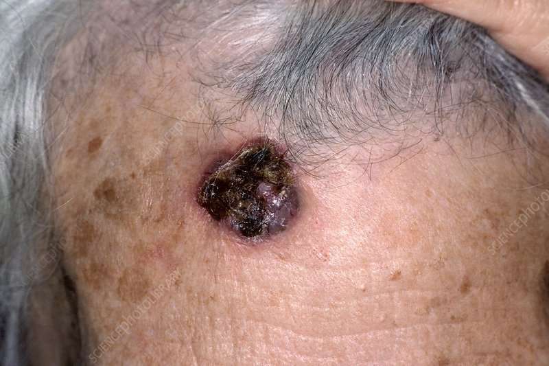
Skin cancers usually present as an abnormal growth on the skin. The growth may have the appearance of a wart, crusty spot, ulcer, mole or sore. It may or may not bleed and can be painful. If you have a preexisting mole, any change in the characteristics of this spot – such as a raised or an irregular border, irregular shape, change in color, increase in size, itching or bleeding – are warning signs of melanoma. Sometimes the first sign of melanoma or squamous cell cancer is an enlarged lymph node.
Johns Hopkins Head and Neck Cancer Surgery Specialists
Our head and neck surgeons and speech language pathologists take a proactive approach to cancer treatment. Meet the Johns Hopkins specialists who will work closely with you during your journey.
Should People Have A Doctor Remove A Dysplastic Nevus Or A Common Mole To Prevent It From Changing Into Melanoma
No. Normally, people do not need to have a dysplastic nevus or common mole removed. One reason is that very few dysplastic nevi or common moles turn into melanoma . Another reason is that even removing all of the moles on the skin would not prevent the development of melanoma because melanoma can develop as a new colored area on the skin . That is why doctors usually remove only a mole that changes or a new colored area on the skin.
Read Also: Can Squamous Cell Carcinoma Metastasis
What Is A Skin Tag
A skin tag is a flesh-colored growth that can be thin and stalky looking or round in shape.
These growths can develop in many areas on your body. Theyre most common in parts where friction is created from skin rubbing. As skin tags age, they may become red or brown in color.
Skin tags are often found in the following areas of the body:
- armpits
I’ve Been Diagnosed With Melanomawhat Happens Next

Doctors use the TNM system developed by the American Joint Committee on Cancer to begin the staging process. Its a classification based on three key factors:
T stands for the extent of the original tumor, its thickness or how deep it has grown and whether it has ulcerated.
What Is Breslow depth?
Breslow depth is a measurement from the surface of the skin to the deepest component of the melanoma.
Tumor thickness: Known as Breslow thickness or Breslow depth, this is a significant factor in predicting how far a melanoma has advanced. In general, a thinner Breslow depth indicates a smaller chance that the tumor has spread and a better outlook for treatment success. The thicker the melanoma measures, the greater its chance of spreading.
Tumor ulceration: Ulceration is a breakdown of the skin on top of the melanoma. Melanomas with ulceration are more serious because they have a greater risk of spreading, so they are staged higher than tumors without ulceration.
N indicates whether or not the cancer has already spread to nearby lymph nodes. The N category also includes in-transit tumors that have spread beyond the primary tumor toward the local lymph nodes but have not yet reached the lymph nodes.
M represents spread or metastasis to distant lymph nodes or skin sites and organs such as the lungs or brain.
After TNM categories are identified, the overall stage number is assigned. A lower stage number means less progression of the disease.
Recommended Reading: Does Insurance Cover Skin Cancer Screening
Diagnosing Squamous Cell Carcinoma
The main way to diagnose squamous cell carcinoma is with a biopsy. This involves having a small piece of tissue removed from the suspicious area and examined in a laboratory.
In the laboratory, a pathologist will examine the tissue under a microscope to determine if it is a skin cancer. He or she will also stage the cancer by the number of abnormal cells, their thickness, and the depth of penetration into the skin. The higher the stage of the tumor, the greater the chance it could spread to other parts of the body.
Squamous cell carcinoma on sun-exposed areas of skin usually does not spread. However, squamous cell carcinoma of the lip, vulva, and penis are more likely to spread. Contact your doctor about any sore in these areas that does not go away after several weeks.
What You Can Do
Check yourself: No matter your risk, examine your skin head-to-toe once a month to identify potential skin cancers early. Take note of existing moles or lesions that grow or change. Learn how to check your skin here.
When in doubt, check it out. Because melanoma can be so dangerous once it advances, follow your instincts and visit your doctor if you see a spot that just doesnt seem right.
Keep in mind that while important, monthly self-exams are not enough. See your dermatologist at least once a year for a professional skin exam.
If youve had a melanoma, follow up regularly with your doctor once treatment is complete. Stick to the schedule your doctor recommends so that you will find any recurrence as early as possible.
Reviewed by:
You May Like: What To Do To Prevent Skin Cancer
Don’t Let Skin Cancer Sneak Up On You
Do you know how to spot skin cancer? In this video, the American Academy of Dermatology used an ultraviolet camera to show people the sun damage hidden underneath their skin. While you cant see all the sun damage on your skin, its important to check the spots you can see before its too late.
Can you spot skin cancer?
Anyone can get skin cancer, regardless of skin color. It is estimated that one in five Americans will develop skin cancer in their lifetime. When caught early, skin cancer is highly treatable.
When Is A Mole A Problem
A mole is a benign growth of melanocytes, cells that gives skin its color. Although very few moles become cancer, abnormal or atypical moles can develop into melanoma over time. “Normal” moles can appear flat or raised or may begin flat and become raised over time. The surface is typically smooth. Moles that may have changed into skin cancer are often irregularly shaped, contain many colors, and are larger than the size of a pencil eraser. Most moles develop in youth or young adulthood. It’s unusual to acquire a mole in the adult years.
Don’t Miss: How To Know You Have Skin Cancer
What Are The Symptoms Of Squamous Cell Cancer With Occult Primary
Key Points Metastatic squamous neck cancer with occult primary is a disease in which squamous cell cancer spreads to lymph nodes in the neck and it is not known where the cancer first formed in the body. Signs and symptoms of metastatic squamous neck cancer with occult primary include a lump or pain in the neck or throat.
What Are The Melanoma Stages And What Do They Mean
Early melanomas
Stage 0 and I are localized, meaning they have not spread.
- Stage 0: Melanoma is localized in the outermost layer of skin and has not advanced deeper. This noninvasive stage is also called melanoma in situ.
- Stage I: The cancer is smaller than 1 mm in Breslow depth, and may or may not be ulcerated. It is localized but invasive, meaning that it has penetrated beneath the top layer into the next layer of skin. Invasive tumors considered stage IA are classified as early and thin if they are not ulcerated and measure less than 0.8 mm.
Find out about treatment options for early melanomas.
Intermediate or high-risk melanomas
Localized but larger tumors may have other traits such as ulceration that put them at high risk of spreading.
- Stage II: Intermediate, high-risk melanomas are tumors deeper than 1 mm that may or may not be ulcerated. Although they are not yet known to have advanced beyond the primary tumor, the risk of spreading is high, and physicians may recommend a sentinel lymph node biopsy to verify whether melanoma cells have spread to the local lymph nodes. Thicker melanomas, greater than 4.0 mm, have a very high risk of spreading, and any ulceration can move the disease into a higher subcategory of stage II. Because of that risk, the doctor may recommend more aggressive treatment.
Learn more about sentinel lymph node biopsy and melanoma treatment options.
Advanced melanomas
Also Check: What Is The Latest Treatment For Melanoma
Tips For Screening Moles For Cancer
Examine your skin on a regular basis. A common location for melanoma in men is on the back, and in women, the lower leg. But check your entire body for moles or suspicious spots once a month. Start at your head and work your way down. Check the “hidden” areas: between fingers and toes, the groin, soles of the feet, the backs of the knees. Check your scalp and neck for moles. Use a handheld mirror or ask a family member to help you look at these areas. Be especially suspicious of a new mole. Take a photo of moles and date it to help you monitor them for change. Pay special attention to moles if you’re a teen, pregnant, or going through menopause, times when your hormones may be surging.
Melanoma Can Be Tricky

Identifying a potential skin cancer is not easy, and not all melanomas follow the rules. Melanomas come in many forms and may display none of the typical warning signs.
Its also important to note that about 20 to 30 percent of melanomas develop in existing moles, while 70 to 80 percent arise on seemingly normal skin.
Amelanotic melanomas are missing the dark pigment melanin that gives most moles their color. Amelanotic melanomas may be pinkish, reddish, white, the color of your skin or even clear and colorless, making them difficult to recognize.
Acral lentiginous melanoma, the most common form of melanoma found in people of color, often appears in hard-to-spot places, including under the fingernails or toenails, on the palms of the hands or soles of the feet.
The takeaway: Be watchful for any new mole or freckle that arises on your skin, a sore or spot that does not heal, any existing mole that starts changing or any spot, mole or lesion that looks unusual.
Acral lentiginous melanoma is the most common melanoma found in people of color.
Recommended Reading: How To Remove Skin Cancer On Face
Diagnosis And Staging What It Means For You
How is melanoma diagnosed?
To diagnose melanoma, a dermatologist biopsies the suspicious tissue and sends it to a lab, where a dermatopathologist determines whether cancer cells are present.
After the disease is diagnosed and the type of melanoma is identified, the next step is for your medical team to identify the stage of the disease. This may require additional tests including imaging such as PET scans, CT scans, MRIs and blood tests.
The stage of melanoma is determined by several factors, including how much the cancer has grown, whether the disease has spread and other considerations. Melanoma staging is complex, but crucial. Knowing the stage helps doctors decide how to best treat your disease and predict your chances of recovery.
For More Information About Skin Cancer
National Cancer Institute, Cancer Information Service Toll-free: 4-CANCER 422-6237TTY : 332-8615
Skin Cancer Foundation
Media file 1: Skin cancer. Malignant melanoma.
Media file 2: Skin cancer. Basal cell carcinoma.
Media file 3: Skin cancer. Superficial spreading melanoma, left breast. Photo courtesy of Susan M. Swetter, MD, Director of Pigmented Lesion and Cutaneous Melanoma Clinic, Assistant Professor, Department of Dermatology, Stanford University Medical Center, Veterans Affairs Palo Alto Health Care System.
Media file 4: Skin cancer. Melanoma on the sole of the foot. Diagnostic punch biopsy site located at the top. Photo courtesy of Susan M. Swetter, MD, Director of Pigmented Lesion and Cutaneous Melanoma Clinic, Assistant Professor, Department of Dermatology, Stanford University Medical Center, Veterans Affairs Palo Alto Health Care System.
Media file 5: Skin cancer. Melanoma, right lower cheek. Photo courtesy of Susan M. Swetter, MD, Director of Pigmented Lesion and Cutaneous Melanoma Clinic, Assistant Professor, Department of Dermatology, Stanford University Medical Center, Veterans Affairs Palo Alto Health Care System.
Continued
Media file 6: Skin cancer. Large sun-induced squamous cell carcinoma on the forehead and temple. Image courtesy of Dr. Glenn Goldman.
Also Check: How Bad Is Squamous Cell Carcinoma
When Melanoma Can’t Be Cured
If your cancer has spread and it is not possible to cure it by surgery, your doctor may still recommend treatment. In this case, treatment may help to relieve symptoms, might make you feel better and may allow you to live longer.Whether or not you choose to have anti-cancer treatment, symptoms can still be controlled. For example, if you have pain, there are effective treatments for this. General practitioners, specialists and palliative care teams in hospitals all play important roles in helping people with cancer.
What Is A Dysplastic Nevus
A dysplastic nevus is a type of mole that looks different from a common mole. A dysplastic nevus may be bigger than a common mole, and its color, surface, and border may be different. It is usually more than 5 millimeters wide . A dysplastic nevus can have a mixture of several colors, from pink to dark brown. Usually, it is flat with a smooth, slightly scaly, or pebbly surface, and it has an irregular edge that may fade into the surrounding skin. Some examples of dysplastic nevi are shown here. More examples are on the What Does a Mole Look Like? page.
Dysplastic Nevi Photos
This dysplastic nevus has a raised area at the center that doctors may call a fried egg appearance.
This dysplastic nevus is more than 5 millimeters in diameter.
This dysplastic nevus is more than 10 millimeters wide .
A dysplastic nevus may occur anywhere on the body, but it is usually seen in areas exposed to the sun, such as on the back. A dysplastic nevus may also appear in areas not exposed to the sun, such as the scalp, breasts, and areas below the waist . Some people have only a couple of dysplastic nevi, but other people have more than 10. People who have dysplastic nevi usually also have an increased number of common moles.
Also Check: Can Melanoma Be Treated Successfully
Dietary Sources Of Vitamin D
The best natural sources of vitamin D in the diet include fatty fish and fish liver oils. Small amounts of vitamin D are also found in egg yolks, beef liver, some mushrooms, ricotta cheese, and some cuts of pork. Vitamin D-fortified foods and beverages provide most of the vitamin D in the U.S. diet. Almost all of the milk in the United States is fortified with vitamin D, and many of the ready-to-eat breakfast cereals provide a small amount of added vitamin D. In addition, specific brands of soy beverages, orange juice, yogurt, margarine, and other foods are also fortified with vitamin D.
Medical Uses of UV Exposure
Dermatologists and other doctors sometimes use UV light to treat health conditions, such as psoriasis, rickets, and eczema. These providers are advised to carefully weigh the risks and benefits of UV treatment for individual patients and carefully monitor doses.,-
Benefits of Being Outdoors
Risks of Indoor Tanning Outweigh Any Potential Benefits
Low levels of sunlight in the winter months may contribute to seasonal affective disorder , and as a result, some indoor tanners may attempt to self-treat SAD with UV exposure through indoor tanning., Medical treatment of SAD frequently incorporates light treatment, but UV wavelengths are not generally recommended .,, In addition, light is thought to affect SAD through the retina, not the skin.
The Most Important Things To Look For Are Skin Growths Or Patches That Are Different From Other Areas Of The Skin And Change Over Time
Features that make a “pimple” highly suspect for skin cancer. Itching and bleeding are other common signs. If you’re treating the the pimple but it’s still isn’t going. When you puncture the pimple’s outer skin, the gunk oozes out. 11.05.2021 · well, it’s just skin cancer, she thought. The most important things to look for are skin growths or patches that are different from other areas of the skin and change over time. 12.06.2019 · these skin cancer pictures will help you differentiate the different types, including basal cell and squamous cell carcinomas, melanoma, actinic keratosis, and merkel cell carcinoma. 17.05.2018 · skin cancer doesn’t mess around. If the bacteria in that gunk splatters and lands inside other pores, it can lead to more pimples. 13.08.2021 · skin cancer can appear as moles, nodules, rashes, scaly patches, or sores that won’t heal. 2 especially, after two months it’s bigger. 3 very firm and hard. “any scab that does not heal normally after one month should be evaluated because normal wounds should heal by that time,” explains estee williams, md, a board certified medical, cosmetic and surgical dermatologist and assistant clinical professor in dermatology at mount sinai.
11.05.2021 · well, it’s just skin cancer, she thought. 1 after a few months it hasn’t budged. 12.06.2019 · pimples can take a long time to go away. If you’re treating the the pimple but it’s still isn’t going. 17.05.2018 · skin cancer doesn’t mess around.
Don’t Miss: How To Protect Your Self From Skin Cancer
How To Tell If A Lump Might Be Cancerous
How they feel Hard, and they don’t hurt or move. You would find one in the lower half of the neck.
Why they pop up The cause of thyroid nodules is not known. After verifying that yours is benign, your M.D. might simply monitor you. If you have additional thyroid symptoms, however, treating the underlying disorder with medication or with radioactive iodine can shrink the lump.
RELATED: Fight Cancer: Make Exercise Your Secret Weapon
How they feel Like a soft grape. They are often tender to the touch. These fluid-filled sacs are common in breasts and the genital area.
Why they pop up Breast cysts tend to wax and wane with your cycle if you have one that persists longer than a month, request an ultrasound or a fine-needle aspiration. Should you find a soft genital bump, it’s likely that a blocked oil duct has caused an epidermoid cyst, says Anita Shivadas, M.D., an internist at the Cleveland Clinic. If it is sensitive, apply warm, moist compresses and antibiotic cream. No pain? Leave it alone.
RELATED: Prevent Cancer: Quit Smoking and Reduce Stress
How they feel Like a squishy ball of tissue that moves easily. These fat deposits show up mostly on the legs, trunk and arms, explains Eileen S. Moore, M.D., assistant professor of medicine at Georgetown University in Washington, D.C.
Why they pop up Lipomas tend to run in families. Unless they are painful or impinge on a nerve or blood vessel, your M.D. can keep an eye on them otherwise, they can be surgically removed.