How Often Should You Check For Skin Cancer
Yearly skin exams are typically recommended as a preventative measure, says Dr. Crutchfield. In addition to a head-to-toe exam, they can also take photos of any suspicious moles.
A monthly skin-check at home is recommended to check for new lesions or to monitor any changes in atypical moles. Do the skin-check by standing naked in front of a full-length mirror, in a room with good lighting, holding a hand mirror, says Dr. King. . Get a friend or partner to do a check of hard to see places like your back.
Bottom line: There are many types of skin cancer, each of which can look different person to personso go see your doc if you notice any marks on your skin that are new or changing or worrisome.
When it comes to reviewing skin cancer pictures and identifying the big C, Dr. Crutchfield’s best advice is “see spot, see spot change, see a dermatologist.”
What To Look For
Because skin cancers appear in many shapes and sizes, its important to know the warning signs associated with basal cell carcinoma , squamous cell carcinoma , melanoma, Merkel cell carcinoma and the precancer actinic keratosis .
If you see something NEW, CHANGING or UNUSUAL, get checked by a dermatologist right away. It could be skin cancer. This includes:
- A growth that increases in size and appears pearly, transparent, tan, brown, black, or multicolored.
- A mole, birthmark or brown spot that increases in size, thickness, changes color or texture, or is bigger than a pencil eraser. Learn the ABCDEs of melanoma.
- A spot or sore that continues to itch, hurt, crust, scab or bleed.
- An open sore that does not heal within three weeks.
Learn more about early detection at TheBigSee.org.
What Are The Signs And Symptoms Of Skin Cancer
Skin cancers are the most commonly diagnosed malignancies in the United States. In fact, more skin cancers will be diagnosed this year than all other types of cancer combined. Advanced skin cancers are easy to identify: because they look very different from other common skin changes.
Advanced skin cancers often present as large, slowly extending, unhealing sores thick, tender or asymptomatic growing tumors or sizable fast-growing black or pink bumps.
However, doctors hope not to have to diagnose these advanced skin cancers. Most skin cancers are easily treatable when diagnosed early, but if identified later, they can cause both a treatment challenge and potentially a significant health risk. To improve treatment outcomes and to reduce health risk, early skin cancer diagnosis is crucial.
The problem is that while advanced skin cancers may be obvious, early skin cancers arent always easy to identify.
“It can be very hard to identify a skin cancer, because hundreds sometimes thousands of harmless skin lesions might look unusual to the untrained eye,” says Gyorgy Paragh, MD, PhD, Chair of the Department of Dermatology at Roswell Park Comprehensive Cancer Center.
When doctors try to identify problem pigmented lesions, they recommend that people look for what they call the ABCDE features. To include the easiest-to-use ugly duckling sign, Dr. Paragh generally recommends extending these features to ABCDEFs.
Read Also: Melanoma Forearm
The Abcdes Of Melanoma
The first five letters of the alphabet are a guide to help you recognize the warning signs of melanoma.
A is for Asymmetry. Most melanomas are asymmetrical. If you draw a line through the middle of the lesion, the two halves dont match, so it looks different from a round to oval and symmetrical common mole.
B is for Border. Melanoma borders tend to be uneven and may have scalloped or notched edges, while common moles tend to have smoother, more even borders.
C is for Color. Multiple colors are a warning sign. While benign moles are usually a single shade of brown, a melanoma may have different shades of brown, tan or black. As it grows, the colors red, white or blue may also appear.
D is for Diameter or Dark. While its ideal to detect a melanoma when it is small, its a warning sign if a lesion is the size of a pencil eraser or larger. Some experts say it is also important to look for any lesion, no matter what size, that is darker than others. Rare, amelanotic melanomas are colorless.
E is for Evolving. Any change in size, shape, color or elevation of a spot on your skin, or any new symptom in it, such as bleeding, itching or crusting, may be a warning sign of melanoma.
If you notice these warning signs, or anything NEW, CHANGING or UNUSUAL on your skin see a dermatologist promptly.
A is for Asymmetry
D is for Diameter or Dark
E is for Evolving
E is for Evolving
Infiltrative Basal Cell Carcinoma

This photo contains content that some people may find graphic or disturbing.
DermNet NZ
Infiltrative basal cell carcinoma occurs when a tumor makes its way into the dermis via thin strands between collagen fibers. This aggressive type of skin cancer is harder to diagnose and treat because of its location. Typically, infiltrative basal cell carcinoma appears as scar tissue or thickening of the skin and requires a biopsy to properly diagnose.
To remove this type of basal cell carcinoma, a specific form of surgery, called Mohs, is used. During a Mohs surgery, also called Mohs micrographic surgery, thin layers of skin are removed until there is no cancer tissue left.
This photo contains content that some people may find graphic or disturbing.
DermNet NZ
Superficial basal cell carcinoma, also known as in situ basal-cell carcinoma, tends to occur on the shoulders or the upper part of the torso, but it can also be found on the legs and arms. This type of cancer isnt generally invasive because it has a slow rate of growth and is fairly easy to spot and diagnose. It appears reddish or pinkish in color and may crust over or ooze. Superficial basal cell carcinoma accounts for roughly 15%-26% of all basal cell carcinoma cases.
Read Also: Melanoma 3c
Grouping Risk Predictors Into Risk Scores
We defined six risk scores by grouping risk factors included within the final 32-factor models . The Demographic risk score includes three factors . The Family history risk score contains only one factor: it is defined as a simple score ranging from 0 to 4, where a value of 4 indicates that the participant reported that his/her father , mother , at least one sibling , and at least one children , developed skin cancer . We explored alternative and more complex definitions, including scores weighted by number of siblings, number of children, or skin cancer-specific family history. However, the simple score out performed these more complex scores at explaining phenotypic variance in the training set. As very few participants reported a score of 4, we combined the scores 3 and 4. The Mole risk score combines four risk factors related to the presence or frequency of moles , and skin conditions . The Susceptibility risk score combines 8 factors related to pigmentation but also skin reaction to sun exposure . The Exposure risk score combines 8 factors that estimate lifetime or current weekly sun exposure . The Miscellaneous risk score combines 7 factors that are not a natural fit in the 5 other risk scores. These risk factors are mainly related to metabolism and personality/behavior .
How To Detect Cancer Early
This article was co-authored by Chris M. Matsko, MD and by wikiHow staff writer, Jessica Gibson. Dr. Chris M. Matsko is a retired physician based in Pittsburgh, Pennsylvania. With over 25 years of medical research experience, Dr. Matsko was awarded the Pittsburgh Cornell University Leadership Award for Excellence. He holds a BS in Nutritional Science from Cornell University and an MD from the Temple University School of Medicine in 2007. Dr. Matsko earned a Research Writing Certification from the American Medical Writers Association in 2016 and a Medical Writing & Editing Certification from the University of Chicago in 2017. This article has been viewed 58,229 times.
If you’ve had family members deal with cancer or you’ve been diagnosed with a precancerous condition, it’s understandable that you might want to be alert for early signs of cancer. Since the signs, severity, and growth of cancer are completely unique to each individual, it’s important to pay attention to any changes in your body. You can also talk with your doctor about doing genetic testing to determine your risk for developing a specific cancer. Being aware of your risks and monitoring potential symptoms can increase your chances of survival if the cancer is detected early.
Also Check: Life Expectancy Metastatic Melanoma
Medical Treatment For Skin Cancer
Surgical removal is the mainstay of therapy for both basal cell and squamous cell carcinomas. For more information, see Surgery.
People who cannot undergo surgery may be treated by external radiation therapy. Radiation therapy is the use of a small beam of radiation targeted at the skin lesion. The radiation kills the abnormal cells and destroys the lesion. Radiation therapy can cause irritation or burning of the surrounding normal skin. It can also cause fatigue. These side effects are temporary. In addition, a topical cream has recently been approved for the treatment of certain low-risk nonmelanoma skin cancers.
In advanced cases, immune therapies, vaccines, or chemotherapy may be used. These treatments are typically offered as clinical trials. Clinical trials are studies of new therapies to see if they can be tolerated and work better than existing therapies.
Syndromes And Genes Associated With A Predisposition For Squamous Cell Carcinoma
Major genes have been defined elsewhere in this summary as genes that are necessary and sufficient for disease, with important pathogenic variants of the gene as causal. The disorders resulting from single-gene pathogenic variants within families lead to a very high risk of disease and are relatively rare. The influence of the environment on the development of disease in individuals with these single-gene disorders is often very difficult to determine because of the rarity of the genetic variant.
Identification of a strong environmental risk factorchronic exposure to UV radiationmakes it difficult to apply genetic causation for SCC of the skin. Although the risk of UV exposure is well known, quantifying its attributable risk to cancer development has proven challenging. In addition, ascertainment of cases of SCC of the skin is not always straightforward. Many registries and other epidemiologic studies do not fully assess the incidence of SCC of the skin owing to: the common practice of treating lesions suspicious for SCC without a diagnostic biopsy, and the relatively low potential for metastasis. Moreover, NMSC is routinely excluded from the major cancer registries such as the Surveillance, Epidemiology, and End Results registry.
With these considerations in mind, the discussion below will address genes associated with disorders that have an increased incidence of skin cancer.
Xeroderma pigmentosum
Multiple self-healing squamous epitheliomata
Oculocutaneous albinism
Don’t Miss: Melanoma On Face Prognosis
Skin Cancer On Scalp Child Idaman
Skin Cancer Scabs On Scalp. Here are a number of highest rated Skin Cancer Scabs On Scalp pictures on internet. We identified it from honorable source. Its submitted by dealing out in the best field. We assume this nice of Skin Cancer Scabs On Scalp graphic could possibly be the most trending topic like we ration it in google gain or facebook.
Subscribe.derbytelegraph.co.uk is an open platform for users to share their favorite wallpapers, By downloading this wallpaper, you agree to our Terms Of Use and Privacy Policy. This image is for personal desktop wallpaper use only, if you are the author and find this image is shared without your permission, DMCA report please Contact Us
I’ve Been Diagnosed With Melanomawhat Happens Next
Doctors use the TNM system developed by the American Joint Committee on Cancer to begin the staging process. Its a classification based on three key factors:
T stands for the extent of the original tumor, its thickness or how deep it has grown and whether it has ulcerated.
What Is Breslow depth?
Breslow depth is a measurement from the surface of the skin to the deepest component of the melanoma.
Tumor thickness: Known as Breslow thickness or Breslow depth, this is a significant factor in predicting how far a melanoma has advanced. In general, a thinner Breslow depth indicates a smaller chance that the tumor has spread and a better outlook for treatment success. The thicker the melanoma measures, the greater its chance of spreading.
Tumor ulceration: Ulceration is a breakdown of the skin on top of the melanoma. Melanomas with ulceration are more serious because they have a greater risk of spreading, so they are staged higher than tumors without ulceration.
N indicates whether or not the cancer has already spread to nearby lymph nodes. The N category also includes in-transit tumors that have spread beyond the primary tumor toward the local lymph nodes but have not yet reached the lymph nodes.
M represents spread or metastasis to distant lymph nodes or skin sites and organs such as the lungs or brain.
After TNM categories are identified, the overall stage number is assigned. A lower stage number means less progression of the disease.
Also Check: How Long Until Melanoma Spreads
From Cats And Dogs To Melanomas And Carcinomas
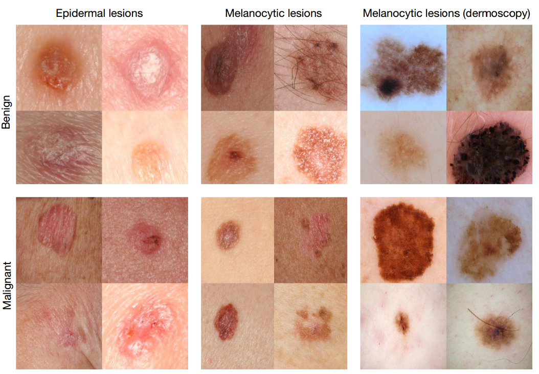
Rather than building an algorithm from scratch, the researchers began with an algorithm developed by Google that was already trained to identify 1.28 million images from 1,000 object categories. While it was primed to be able to differentiate cats from dogs, the researchers needed it to know a malignant carcinoma from a benign seborrheic keratosis.
Theres no huge dataset of skin cancer that we can just train our algorithms on, so we had to make our own, said Brett Kuprel, co-lead author of the paper and a graduate student in the Thrun lab. We gathered images from the internet and worked with the medical school to create a nice taxonomy out of data that was very messy the labels alone were in several languages, including German, Arabic and Latin.
After going through the necessary translations, the researchers collaborated with dermatologists at Stanford Medicine, as well as Helen M. Blau, professor of microbiology and immunology at Stanford and co-author of the paper. Together, this interdisciplinary team worked to classify the hodgepodge of internet images. Many of these, unlike those taken by medical professionals, were varied in terms of angle, zoom and lighting. In the end, they amassed about 130,000 images of skin lesions representing over 2,000 different diseases.
Brett Kuprel
Risk Factors For Squamous Cell Carcinoma

Sun exposure and other risk factors
Sun exposure is the major known environmental factor associated with the development of skin cancer of all types however, different patterns of sun exposure are associated with each major type of skin cancer. Unlike basal cell carcinoma , SCC is associated with chronic exposure, rather than intermittent intense exposure to ultraviolet radiation. Occupational exposure is the characteristic pattern of sun exposure linked with SCC. Other agents and factors associated with SCC risk include tanning beds, arsenic, therapeutic radiation , chronic skin ulceration, and immunosuppression.
Characteristics of the skin
Like melanoma and BCC, SCC occurs more frequently in individuals with lighter skin than in those with darker skin. A case-control study of 415 cases and 415 controls showed similar findings relative to Fitzpatrick type I skin, individuals with increasingly darker skin had decreased risks of skin cancer . The same study found that blue eyes and blond/red hair were also associated with increased risks of SCC, with crude ORs of 1.7 for blue eyes, 1.5 for blond hair, and 2.2 for red hair.
Immunosuppression
Personal history of BCC, SCC, and melanoma skin cancers
Family history of squamous cell carcinoma or associated premalignant lesions
Read Also: Signs Of Stage 4 Cancer
Does Skin Cancer Affect People With Skin Of Color
People of all skin tones can develop skin cancer. If you are a person of color, you may be less likely to get skin cancer because you have more of the brown pigment, melanin, in your skin.
Although less prevalent than in nonwhite people, when skin cancer does develop in people of color, its often found late and has a worse prognosis. If youre Hispanic, the incidence of melanoma has risen by 20% in the past two decades. If youre Black and develop melanoma, your five-year survival rate is 25% lower than it is for white people . Part of the reason may be that it develops in less typical, less sun-exposed areas and its often in late-stage when diagnosed.
How Are Moles Evaluated
If you find a mole or spot that has any ABCDE’s of melanoma — or one that’s tender, itching, oozing, scaly, doesn’t heal or has redness or swelling beyond the mole — see a doctor. Your doctor may want to remove a tissue sample from the mole and biopsy it. If found to be cancerous, the entire mole and a rim of normal skin around it will be removed and the wound stitched closed. Additional treatment may be needed.
Also Check: Stage 3 Melanoma Life Expectancy
Design Setting And Participants
A multicenter retrospective cohort study with a case note review of consecutive secondary care consultations was conducted using data from 2 urgent suspected skin cancer screening clinics in UK National Health Service trusts. The study was performed from January 1, 2015, to March 31, 2016, and data analysis was performed from October 14, 2018, to February 1, 2019. Patients included those presenting with a skin lesion suspicious of malignancy who were referred to the urgent suspected skin cancer clinic over 15 months. Patients who accepted and received a TBSE were subsequently included in the analysis.
Something Just Looks A Little Odd
Your skin is always changing in fact, it regenerates itself all the time. So, if you notice a spot that doesnt go away over the course of a month, it means that this spot sits in the lower layers of your skin. These skin abnormalities should be checked out by a doctor.
It is a good idea to keep track of the size and shape of your moles so that you can show your doctor a timeline to help with diagnosis. SkinVision is ideal for this, as it enables you to detect signs of skin cancer in time and allows you to archive photos of your moles to track any possible changes.
You May Like: Invasive Ductal Carcinoma Stage 2 Survival Rate