Fine Needle Aspiration Biopsy
FNA biopsy is not used on suspicious moles. But it may be used, for example, to biopsy large lymph nodes near a melanoma to find out if the melanoma has spread to them.
For this type of biopsy, the doctor uses a syringe with a thin, hollow needle to remove very small pieces of a lymph node or tumor. The needle is smaller than the needle used for a blood test. A local anesthetic is sometimes used to numb the area first. This test rarely causes much discomfort and does not leave a scar.
If the lymph node is just under the skin, the doctor can often feel it well enough to guide the needle into it. For a suspicious lymph node deeper in the body or a tumor in an organ such as the lung or liver, an imaging test such as ultrasound or a CT scan is often used to help guide the needle into place.
FNA biopsies are not as invasive as some other types of biopsies, but they may not always collect enough of a sample to tell if a suspicious area is melanoma. In these cases, a more invasive type of biopsy may be needed.
Research Is Promising But Accuracy Isnt Quite There
Of all the apps discussed here, SkinVision seems to have the most research behind it.
A 2014 study on an older version of SkinVision reported 81% accuracy in detecting melanoma, which at the time researchers said was insufcient to detect melanoma accurately.
However, a new 2019 study published in the Journal of the European Academy of Dermatology and Venereology determined that SkinVision can detect 95% of skin cancer cases. Its encouraging to see the company continue to work on app accuracy, as early detection of skin cancer is the number-one way to achieve successful treatment.
In another study, researchers from the University of Pittsburgh, analyzed four smartphone apps that claim to detect skin cancer. We dont know the exact apps, as theyre named only as Application 1, 2, 3 and 4. Three of the apps used algorithms to send immediate feedback about the persons risk of skin cancer, and the fourth app sent the photos to a dermatologist.
Unsurprisingly, the researchers found the fourth app be the most accurate. The other three apps were found to incorrectly categorize a large number of skin lesions, with one missing nearly 30% of melanomas, classifying them as low-risk lesions.
A 2018 Cochrane review of prior research found that AI-based skin cancer detection has not yet demonstrated sufficient promise in terms of accuracy, and they are associated with a high likelihood of missing melanomas.
Who Is At Risk
People with fair skin and lighter eyes and hair tend to be particularlyvulnerable to skin cancer. Other risk factors include a family history ofmelanoma, more time spent unprotected in the sun, early childhoodsunburns, immunosuppressive disorders, a weakened immune system, and havingmany freckles or moles.
Both men and women are at risk, but there is one troublingtrend: an alarming surge in melanoma rates in young women.This is largely due to tanning from the sun and in tanning salons. Tanning either at beaches or salons is a major risk factor forskin cancers.
Telemedicine Dermatology Services
Recommended Reading: Basal Cell Carcinoma Syndrome
What Are The Signs Of Skin Cancer
The most common warning sign of skin cancer is a change on your skin, typically a new growth, or a change in an existing growth or mole. The signs and symptoms of common and less common types of skin cancers are described below.
Basal cell carcinoma
Basal cell cancer is most commonly seen on sun-exposed areas of skin including your hands, face, arms, legs, ears, mouths, and even bald spots on the top of your head. Basal cell cancer is the most common type of skin cancer in the world. In most people, its slow growing, usually doesnt spread to other parts of the body and is not life-threatening.
Signs and symptoms of basal cell carcinoma include:
- A small, smooth, pearly or waxy bump on the face, ears, and neck.
- A flat, pink/red- or brown-colored lesion on the trunk or arms and legs.
- Areas on the skin that look like scars.
- Sores that look crusty, have a depression in the middle or bleed often.
Squamous cell carcinoma
Squamous cell cancer is most commonly seen on sun-exposed areas of skin including your hands, face, arms, legs, ears, mouths, and even bald spots on the top of your head. This skin cancer can also form in areas such as mucus membranes and genitals.
Signs and symptoms of squamous cell carcinoma include:
- A firm pink or red nodule.
- A rough, scaly lesion that might itch, bleed and become crusty.
Melanoma
Signs and symptoms of melanoma include:
- A brown-pigmented patch or bump.
- A mole that changes in color, size or that bleeds.
Looking For Signs Of Skin Cancer

Non melanoma skin cancers tend to develop most often on skin thats exposed to the sun.
To spot skin cancers early it helps to know how your skin normally looks. That way, youll notice any changes more easily.
To look at areas you cant see easily, you could try using a hand held mirror and reflect your skin onto another mirror. Or you could get your partner or a friend to look. This is very important if youre regularly outside in the sun for work or leisure.
You can take a photo of anything that doesnt look quite right. If you can its a good idea to put a ruler or tape measure next to the abnormal area when you take the photo. This gives you a more accurate idea about its size and can help you tell if its changing. You can then show these pictures to your doctor.
Recommended Reading: Can You Have Cancer Without A Tumor
How Can I Prevent Skin Cancer
For all types of skin cancer, the first lines of defense are awareness and prevention. Prevention steps center on avoiding ultraviolet radiation exposure from both sunlight and tanning beds. This means staying out of the sun, especially when the suns rays are strongest, between 11 a.m. and 3 p.m. using a broad-spectrum water-resistant sunscreen with SPF of at least 30 and covering exposed skin with protective clothing when outdoors, even on a cloudy day.
Perform a skin self-exam
Know What Youre Looking For
The deadliest form of skin cancer is known as melanoma. Its critical to identify melanoma as early as possible. This can be done by performing self-examinations regularly. To spot both melanoma and non-melanoma skin cancers, youll need to take note of any new moles or growths on your body.
Also, remember to keep an eye on existing moles or growths that begin to grow or change in any way. Some more obvious red flags include any lesions that start to change, itch, or bleed and never seem to heal. Read on for more information about how to spot skin cancer.
Also Check: Stage 3 Invasive Ductal Carcinoma Survival Rate
What Do The Results Mean
If a mole or other mark on your skin looks like it might be a sign of cancer, your provider will probably order another test, called a skin biopsy, to make a diagnosis. A skin biopsy is a procedure that removes a small sample of skin for testing. The skin sample is looked at under a microscope to check for cancer cells. If you are diagnosed with skin cancer, you can begin treatment. Finding and treating cancer early may help prevent the disease from spreading.
First Of All What Is A Mole
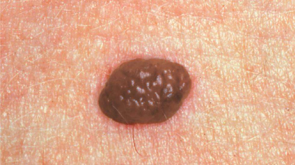
A mole or nevus is a dark spot on our skin comprised of skin cells that have grown in a group rather than individually. These cells are called melanocytes and are responsible for producing melanin, the pigment in our skin. Moles appear on our skin from sun exposure , or we are born with them. Although the number of moles varies from person to person, fair-skinned people generally have more moles due to lower amounts of melanin in their skin. The average number of moles for adults is between 10 and 40. Moles can even come and go with hormonal changes such as pregnancy or puberty.Most people develop more moles on their skin naturally with age and sun exposure, and usually, these moles are harmless. However, we need to conduct regular skin checks to check whether anything has changed.
Don’t Miss: Invasive Ductal Carcinoma Grade 3 Survival Rate
What Is The Outlook For People With Skin Cancer
Nearly all skin cancers can be cured if they are treated before they have a chance to spread. The earlier skin cancer is found and removed, the better your chance for a full recovery. Ninety percent of those with basal cell skin cancer are cured. It is important to continue following up with a dermatologist to make sure cancer does not return. If something seems wrong, call your doctor right away.
Most skin cancer deaths are from melanoma. If you are diagnosed with melanoma:
- The five-year survival rate if its detected before it spreads to the lymph nodes is 99%.
- The five-year survival rate if it has spread to nearby lymph nodes is 66%.
- The five-year survival rate if it has spread to distant lymph nodes and other organs is 27%.
Warning Signs Of Skin Cancer Moles
It is not uncommon for a mole to change gradually over time. Some moles may become darker or flatten as you age. However, you should take note if a mole changes rapidly. Quick changes can be a sign that something isnt right. You should also keep an eye on moles that appear during adulthood as they are more likely to pose a risk.
Atypical moles, also known as dysplastic nevi, tend to be larger than common moles, have irregular edges, and be uneven in color. An atypical mole isnt always a cancerous or pre-cancerous mole, but if you have any, you are advised to have them checked by your doctor to exclude any risks.
Look for these warning signs of skin cancer moles:
- A change in size
- A change in shape
- A change in color
- A loss of symmetry
- Itchiness, pain or bleeding
- Crustiness
- Exhibiting three different shades of brown or black
- A change in elevation
If you notice any of these symptoms, contact a doctor for a skin check.
View an overview of skin cancer images
Read Also: Skin Cancer Pictures Mayo Clinic
Checking Yourself And Your Loved Ones
Start by checking your entire body, including skin not normally exposed to the sun. You could ask for help from someone else to check difficult-to-see areas, such as your back, neck and scalp.
We recommend that you follow the Ugly Duckling rule. The idea behind the Ugly Duckling rule is that you compare your moles with each other. If any mole stands out or looks different from that of nearby moles, it is the ugly duckling, and we advise you contact a doctor to get an expert opinion.
Skin Cancer Diagnosis Always Requires A Skin Biopsy

When you see a dermatologist because youve found a spot that might be skin cancer, your dermatologist will examine the spot.
If the spot looks like it could be a skin cancer, your dermatologist will remove it all or part of it. This can easily be done during your appointment. The procedure that your dermatologist uses to remove the spot is called a skin biopsy.
Having a skin biopsy is essential. Its the only way to know whether you have skin cancer. Theres no other way to know for sure.
What your dermatologist removes will be looked at under a microscope. The doctor who examines the removed skin will look for cancer cells. If cancer cells are found, your biopsy report will tell you what type of skin cancer cells were found. When cancer cells arent found, your biopsy report will explain what was seen under the microscope.
You May Like: Lobular Breast Cancer Survival Rates
I’ve Been Diagnosed With Melanomawhat Happens Next
Doctors use the TNM system developed by the American Joint Committee on Cancer to begin the staging process. Its a classification based on three key factors:
T stands for the extent of the original tumor, its thickness or how deep it has grown and whether it has ulcerated.
What Is Breslow depth?
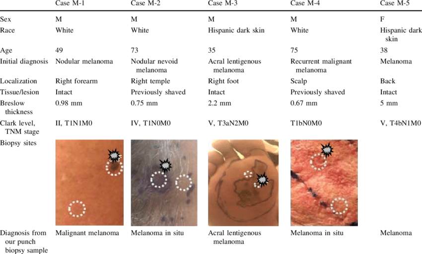
Breslow depth is a measurement from the surface of the skin to the deepest component of the melanoma.
Tumor thickness: Known as Breslow thickness or Breslow depth, this is a significant factor in predicting how far a melanoma has advanced. In general, a thinner Breslow depth indicates a smaller chance that the tumor has spread and a better outlook for treatment success. The thicker the melanoma measures, the greater its chance of spreading.
Tumor ulceration: Ulceration is a breakdown of the skin on top of the melanoma. Melanomas with ulceration are more serious because they have a greater risk of spreading, so they are staged higher than tumors without ulceration.
N indicates whether or not the cancer has already spread to nearby lymph nodes. The N category also includes in-transit tumors that have spread beyond the primary tumor toward the local lymph nodes but have not yet reached the lymph nodes.
M represents spread or metastasis to distant lymph nodes or skin sites and organs such as the lungs or brain.
After TNM categories are identified, the overall stage number is assigned. A lower stage number means less progression of the disease.
When Should I See My Doctor
Its important to check your own skin regularly to find any new or changing spots.
See your doctor or dermatologist straight away if you notice any changes to your skin, such as:
- an ‘ugly duckling’ a spot that looks or feels different to any others
- a spot that changes size, shape, colour or texture over time
- a sore that doesnt go away after a few weeks
- a sore that itches or bleeds
See the ‘ABCDE’ of skin cancer, above.
Read Also: Large Cell Cancers
The Abcdes Of Melanoma
The first five letters of the alphabet are a guide to help you recognize the warning signs of melanoma.
A is for Asymmetry. Most melanomas are asymmetrical. If you draw a line through the middle of the lesion, the two halves dont match, so it looks different from a round to oval and symmetrical common mole.
B is for Border. Melanoma borders tend to be uneven and may have scalloped or notched edges, while common moles tend to have smoother, more even borders.
C is for Color. Multiple colors are a warning sign. While benign moles are usually a single shade of brown, a melanoma may have different shades of brown, tan or black. As it grows, the colors red, white or blue may also appear.
D is for Diameter or Dark. While its ideal to detect a melanoma when it is small, its a warning sign if a lesion is the size of a pencil eraser or larger. Some experts say it is also important to look for any lesion, no matter what size, that is darker than others. Rare, amelanotic melanomas are colorless.
E is for Evolving. Any change in size, shape, color or elevation of a spot on your skin, or any new symptom in it, such as bleeding, itching or crusting, may be a warning sign of melanoma.
If you notice these warning signs, or anything NEW, CHANGING or UNUSUAL on your skin see a dermatologist promptly.
A is for Asymmetry
D is for Diameter or Dark
E is for Evolving
E is for Evolving
How Is Skin Cancer Treated
Treatment depends upon the stage of cancer. Stages of skin cancer range from stage 0 to stage IV. The higher the number, the more cancer has spread.
Sometimes a biopsy alone can remove all the cancer tissue if the cancer is small and limited to your skins surface only. Other common skin cancer treatments, used alone or in combination, include:
Cryotherapy uses liquid nitrogen to freeze skin cancer. The dead cells slough off after treatment. Precancerous skin lesions, called actinic keratosis, and other small, early cancers limited to the skins top layer can be treated with this method.
Excisional surgery
This surgery involves removing the tumor and some surrounding healthy skin to be sure all cancer has been removed.
Mohs surgery
With this procedure, the visible, raised area of the tumor is removed first. Then your surgeon uses a scalpel to remove a thin layer of skin cancer cells. The layer is examined under a microscope immediately after removal. Additional layers of tissue continue to be removed, one layer at a time, until no more cancer cells are seen under the microscope.
Mohs surgery removes only diseased tissue, saving as much surrounding normal tissue as possible. Its most often used to treat basal cell and squamous cell cancers and near sensitive or cosmetically important areas, such as eyelids, ears, lips, forehead, scalp, fingers or genital area.
Curettage and electrodesiccation
Chemotherapy and immunotherapy
You May Like: Metastatic Melanoma Cancer Life Expectancy
Are There Complications Of Skin Cancer Treatment
Most skin cancer treatments involve some localised damage to surrounding healthy skin such as swelling, reddening or blistering of the skin where the cancer is removed. Your doctor will explain any specific risks, which may include:
- pain or itching where the skin has been treated, or if lymph nodes have been removed
- scarring or changes to skin colour, after a skin cancer has been removed
- bleeding during or after surgery for more complicated skin cancers
- reactions sometimes your body may react to medicines used in treatment or surgery
- lymphoedema if your lymph nodes have been removed your neck, arm or leg may swell with fluid.
Its best to manage complications as early as possible, so ask your doctor for advice.