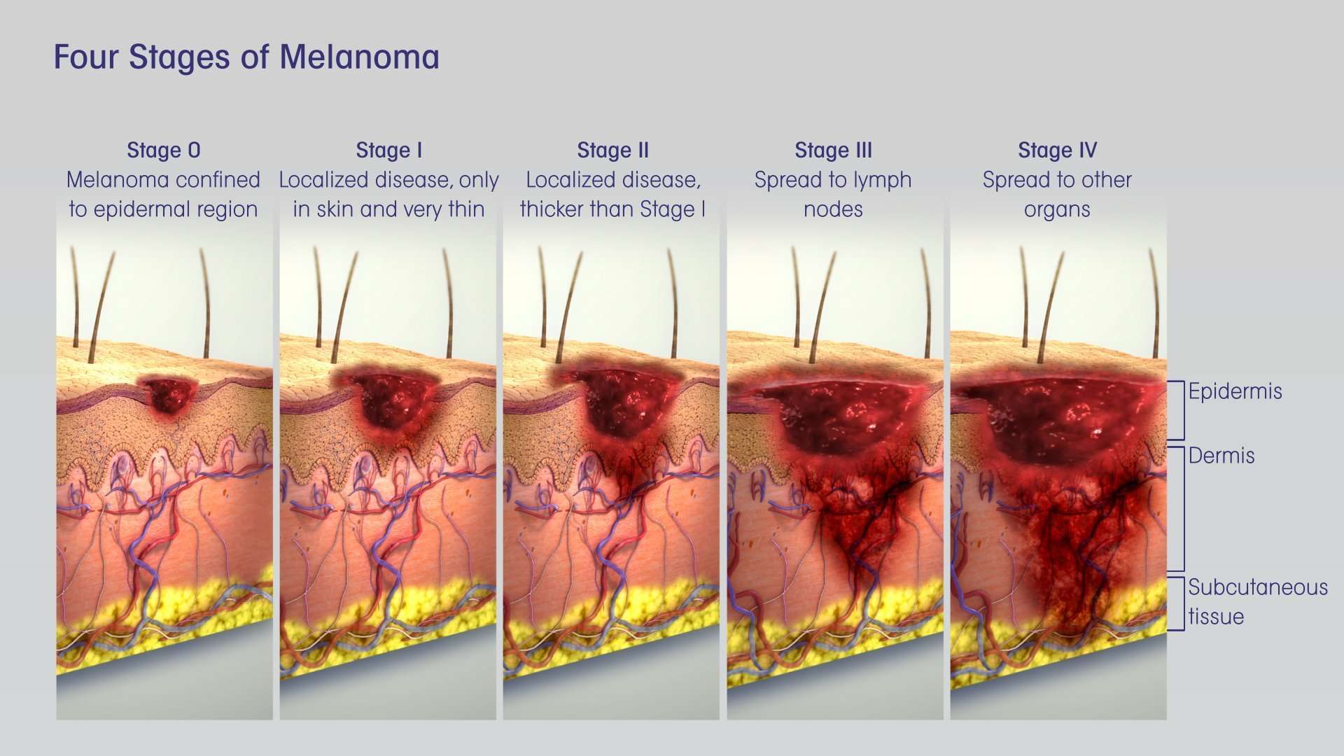
The Five Stages Of Skin Cancer
Cancer in the skin thats at high risk for spreading shares features with basal cell carcinoma and squamous cell carcinoma. Some of these features are:
- Not less than 2 mm in thickness
- Has spread into the inner layers of the skin
- Has invaded skin nerves
Stage 0
In the earliest stage, cancer is only present in the upper layer of the skin. You may notice the appearance of blood vessels or a dent in the center of the skin growth. There are no traces of malignant cells beyond this layer.
Stage 1
At stage 1, cancer has not spread to muscles, bone, and other organs. It measures roughly 4/5 of an inch. Theres a possibility that it may have spread into the inner layer of the skin.
Stage 2
In this stage, cancer has become larger than 4/5 of an inch. Cancer still has not spread to muscles, bone, and other organs.
Stage 3
At stage 3, the cancer is still larger than 4/5 of an inch. Facial bones or a nearby lymph node may have been affected, but other organs remain safe. It may also spread to areas below the skin, such as into muscle, bone, and cartilage but not far from the original site.
Stage 4
Cancer can now be of any size and has likely spread into lymph nodes, bones, cartilage, muscle, or other organs. Distant organs such as the brain or lungs may also be affected. In rare cases, this stage might cause death when allowed to grow and become more invasive.
What Tests Are Used To Stage Melanoma
There are several tests your doctor can use to stage your melanoma. Your doctor may use these tests:
- Sentinel Lymph Node Biopsy: Patients with melanomas deeper than 0.8 mm, those who have ulceration under the microscope in tumors of any size or other less common concerning features under the microscope, may need a biopsy of sentinel lymph nodes to determine if the melanoma has spread. Patients diagnosed via a sentinel lymph node biopsy have higher survival rates than those diagnosed with melanoma in lymph nodes via physical exam.
- Computed Tomography scan: A CT scan can show if melanoma is in your internal organs.
- Magnetic Resonance Imaging scan: An MRI scan is used to check for melanoma tumors in the brain or spinal cord.
- Positron Emission Tomography scan: A PET scan can check for melanoma in lymph nodes and other parts of your body distant from the original melanoma skin spot.
- Blood work: Blood tests may be used to measure lactate dehydrogenase before treatment. Other tests include blood chemistry levels and blood cell counts.
Factors Used For Staging Melanoma
To determine the stage of a melanoma, the lesion and some surrounding healthy tissue need to be surgically removed and analyzed using a microscope. Doctors use the melanomas thickness, measured in millimeters , and the other characteristics described in Diagnosis to help determine the diseases stage.
Doctors also use results from diagnostic tests to answer these questions about the stage of melanoma:
-
How thick or deep is the original melanoma, often called the primary melanoma or primary tumor?
-
Where is the melanoma located?
-
Has the melanoma spread to the lymph nodes? If so, where and how many?
-
Has the melanoma metastasized to other parts of the body? If so, where and how much?
The results are combined to determine the stage of melanoma for each person. The stages of melanoma include: stage 0 and stages I through IV . The stage provides a common way of describing the cancer, so doctors can work together to create the best treatment plan and understand a patients prognosis.
You May Like: What Is The Survival Rate For Invasive Ductal Carcinoma
Curettage Electrodesiccation And Cryotherapy
Some dermatologists perform curettage, electrodesiccation, and cryotherapy to treat skin cancer. These are considered to be destructive techniques that are best suited for small, superficial carcinomas with definite borders. During the procedure, layers of skin cells are scraped away using a curette. Any remaining cancer cells are destroyed with the use of an electric needle.
In some cases, liquid nitrogen or cryotherapy is used to freeze the margins of the treatment area. Extremely low temperatures kill the malignant skin cells and create a wound, which will heal in a few weeks. The treatment may leave scars that are flat and round, similar to the size of the skin cancer lesion.
What Is Cancer Staging

Staging is the process of measuring how far a cancer has spread when it is first diagnosed. It often involves having scans, biopsies and other tests.
Knowing the stage of a cancer is important as it helps doctors to work out the best treatment options.
It also means the person with cancer can fully understand their situation and discuss any concerns they have.
There are different staging systems for different cancers, but they generally use either the:
Also Check: Large Cell Carcinoma Definition
How Common Is Subungual Melanoma
Subungual melanoma is a very infrequent cancer, accounting for approximately 5% of all cases of melanoma. Usually, it is located on the thumb and the big toe. The average age of people affected by this type of cancer is between 60 and 70 years old. Then, subungual melanoma is a rare pathology that, due to a late diagnosis, it is complex to predict. Sometimes it is confused with a subungual hematoma, which is a common trivial pathology. Therefore, subungual melanoma is the most common type of melanoma diagnosed in highly pigmented individuals.
The Warning Signs Of Skin Cancer
Skin cancers including melanoma, basal cell carcinoma, and squamous cell carcinoma often start as changes to your skin. They can be new growths or precancerous lesions changes that are not cancer but could become cancer over time. An estimated 40% to 50% of fair-skinned people who live to be 65 will develop at least one skin cancer. Learn to spot the early warning signs. Skin cancer can be cured if its found and treated early.
Read Also: Malignant Breast Cancer Survival Rate
Basal Cell Carcinoma Staging
Staging is the process of determining whether cancer has spread and, if so, how far. The stage of the disease may affect the treatment plan.
The stage is based on the size of the tumor, how deeply into the skin it has grown, and whether cancer has spread beyond the tumor to the lymph nodes. Your doctor will look at the results of the biopsy to determine the stage. In rare cases, your doctor may recommend imaging such as CT or PET-CT scan to see if the cancer has spread beyond the skin
Stages are numbered in Roman numerals between 0 and IV.
Most non-melanoma skin cancers are Stage 0 or Stage 1. Stage 3 and 4 are relatively rare. Based on the type of cancer, the stage of cancer, your overall health, and other factors, your doctor works with you to develop a treatment plan.
High risk features for primary tumor staging
- Depth/invasion: > 2 mm thickness , Clark level IV, Perineural invasion
- Anatomic: Primary site ear
- Location: Primary site hair-bearing lip
- Differentiation: Poorly differentiated or undifferentiated
Stage 3 Peritoneal Mesothelioma
Peritoneal mesothelioma is the second-most common form of the disease. Instead of a formal staging system to measure progression, physicians typically use the existing Peritoneal Cancer Index to grade tumors in the abdomen. In addition, the PCI helps doctors determine the stage in many other abdominal cancers.
The PCI ranges from 0 to 39, measuring the spread of tumors across 13 different abdominal sectors. A score between 21 and 30 indicates stage 3 peritoneal mesothelioma. The characteristics of this stage are tumors localized within the abdomen, with some spread to nearby lymph nodes.
If a doctor refers to peritoneal mesothelioma as stage 3, it usually means tumors have spread throughout the abdominal lining and to nearby lymph nodes.Dr. Daniel A. LandauOncologist and hematologist
Also Check: What Are The Early Stages Of Melanoma
Don’t Miss: Basal Cell Carcinoma Etiology
How Fast Does Melanoma Spread
Melanoma is a deadly form of skin cancer because of its ability to metastasize to local lymph nodes and other organs. It is estimated that melanoma kills, on average, over 10,000 people in the United States every year.
The first sign of flat melanoma is usually a new spot or an existing mole or freckle that changes in appearance. Some changes can include:
- A spot that has grown in size
- A spot where the edges are looking irregular versus smooth and even
- A spot that has a range of colors such as brown, black, blue, red, white or light gray.
- A spot that has become itchy or is bleeding
According to Dr. Andrew Duncanson, board-certified dermatologist at Forefront Dermatology, It is important to know that melanoma can appear on areas of the skin not normally exposed to the sun such as under the arm, chest, and buttocks. It can also appear in areas that you are not able to see easily on your own including the ears, scalp, back of legs, and bottom of feet. I always recommend to my patients to look for the ugly duckling spot the new spot that doesnt look like any others. Additionally, ask a family member to look over the hard to see areas monthly, while also getting an annual skin cancer exam by a board-certified dermatologist to detect skin cancer early.
Stage I And Stage Ii Melanoma
Stage I and stage II melanoma describe invasive cancer that has grown below the epidermis to the next layer of skin, the dermis. It has not reached the lymph nodes.
Two major factors help determine the seriousness of stage I melanoma and stage II melanoma: Breslow depth and ulceration.
Breslow depth is a measurement that doctors use to describe the depth of an invasive melanoma in millimeters. It measures how far melanoma cells have reached below the surface of the skin. The thinner the melanoma, the better the chances for a cure.
Ulceration means that there is broken skin covering the melanoma. This breakage can be so small that it can only be seen under a microscope. Ulceration is an important factor in staging. A melanoma with ulceration may require more aggressive treatment than a melanoma of the same size without ulceration.
Melanoma is considered stage 1A when:
- the tumor is less than or equal to 1 millimeter thick in Breslow depth
Melanoma is considered stage IB when:
- the tumor is 1.1 to 2 millimeters thick in Breslow depth without ulceration
Melanoma is considered stage IIA when:
- the tumor is 1.1 to 2 millimeters thick in Breslow depth with ulceration
- the tumor is 2.1 to 4 millimeters thick in Breslow depth without ulceration
Melanoma is considered stage IIB when:
- the tumor is 2.1 to 4 millimeters thick in Breslow depth with ulceration
- the tumor is more than 4 millimeters in Breslow depth without ulceration
Melanoma is considered stage IIC when:
Don’t Miss: Does Skin Cancer Itch And Burn
Treatments For Stage Ii Melanoma
As with stage I, stage II melanoma is typically treated with wide excision surgery, which cuts out the melanoma along with a margin of healthy surrounding skin. In the case of stage II melanoma, many doctors will recommend looking for cancer in nearby lymph nodes by performing a sentinel lymph node biopsy, which may necessitate further treatment if cancer cells are found.
Biological Therapies And Melanoma

Biological therapies are treatments using substances made naturally by the body. Some of these treatments are called immunotherapy because they help the immune system fight the cancer, or they occur naturally as part of the immune system. There are many biological therapies being researched and trialled, which in the future may help treat people with melanoma. They include monoclonal antibodies and vaccine therapy.
Don’t Miss: What Is Braf Testing In Melanoma
Can You Have Melanoma For Years And Not Know
How long can you have melanoma and not know it? It depends on the type of melanoma. For example, nodular melanoma grows rapidly over a matter of weeks, while a radial melanoma can slowly spread over the span of a decade. Like a cavity, a melanoma may grow for years before producing any significant symptoms.
Squamous Cell Skin Cancer
This is the second most common form of skin cancer, it occurs most commonly on the head and neck, and exposed arms. However, these are frequently seen on the front of the legs as well, or the shin area. This form of skin cancer grows more quickly, and though it can be confined to the top layer of skin, it frequently grows roots. Squamous cell carcinoma can be more aggressive and does have a potential to spread internally. This is more likely in cases where an individual is immunosuppressed, or the tumor is invading deeply in the second layer of skin, or tracking along nerves. These tumors need to be treated early as they are not only locally destructive, but can spread along nerves, into lymph nodes, and internally.
Recommended Reading: What Is The Survival Rate For Invasive Ductal Carcinoma
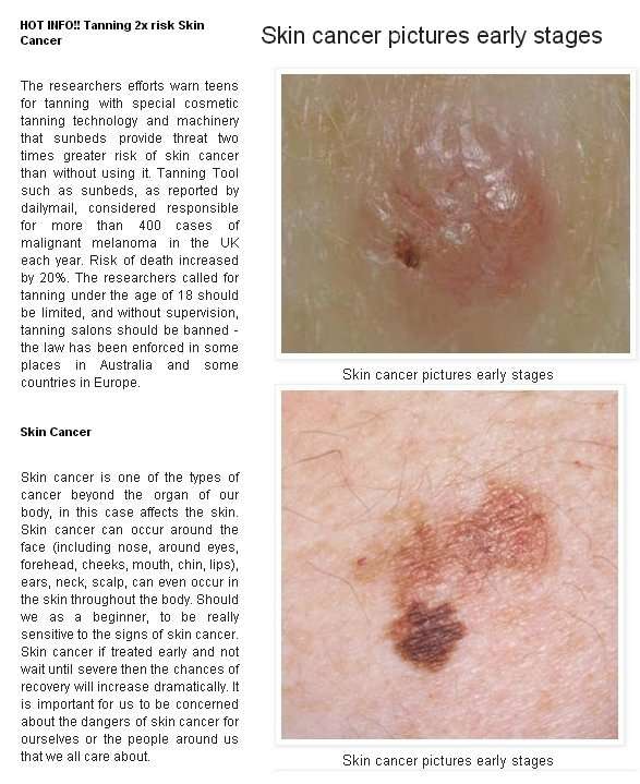
Skin Cancer Pictures By Type
Skin cancer is the most common form of cancer. There are several different types of skin cancer with Basal Cell Carcinoma, Squamous Cell Carcinoma, Bowens Disease, Keratoacanthoma, Actinic Keratosis and Melanoma most commonly occurring.
Basal cell carcinoma is the most common form of skin cancer, and least dangerous whereas melanoma is the most dangerous type.
Below you will find skin cancer pictures of these six types, but remember that skin cancer should be diagnosed by a doctor. Comparing your skin lesion to skin cancer images found online cannot replace medical examination.
If you have any pigmented mole or non-pigmented mark on your skin that looks different from the other marks or moles on your skin, that is new or that has undergone change, is bleeding or wont heal, is itching or in any way just seems off, visit your doctor without delay dont lose time comparing your mole or mark with various pictures of skin cancer.
If you want to be proactive about your health, you may want to photograph areas of your skin routinely including individual moles or marks to familiarise yourself with the appearance of your skin . A skin monitoring app may be a useful tool to assist in that process.
MIISKIN PROMO
What Are The Stages Of Melanoma
Cancerstaging is how doctors describe the extent of cancer in your body. Staging is defined by the characteristics of the original melanomatumor and if/how far it has spread in your body.
Melanoma is divided into stages using five Roman numerals and up to four letters that indicate a higher risk within each stage. The stage is determined mostly by specific details about the tumor and its growth that are tallied in a system called TNM. Read more about the TNM system.
Your stage is important because cancer treatment options and prognoses are determined by stage.
Also Check: Osteomyoma
Less Common Types Of Skin Cancer
Kaposi sarcoma
This is a rare form of skin cancer that develops in the skins blood vessels and causes red or purple patches. It often attacks people with weakened immune systems, such as individuals with AIDS, or in people taking medications that suppress their immune system, such as patients whove received organ transplants.
Merkel cell carcinoma
Merkel cell carcinoma causes firm, shiny nodules that occur on the surface or just beneath the skin and in hair follicles. Merkel cell carcinoma most often appears on the head, neck and torso.
Sebaceous gland carcinoma
This rare but aggressive cancer develops in the skins oil glands. Sebaceous gland carcinomas which usually appear as hard, painless nodules can develop anywhere, but frequently occur on the eyelid, where they can be mistaken for other eyelid problems.
Tests Or Procedures That Examine The Skin Are Used To Diagnose Basal Cell Carcinoma And Squamous Cell Carcinoma Of The Skin
The following procedures may be used:
- Physical exam and health history: An exam of the body to check general signs of health, including checking for signs of disease, such as lumps or anything else that seems unusual. A history of the patients health habits and past illnesses and treatments will also be taken.
- Skin exam: An exam of the skin for bumps or spots that look abnormal in color, size, shape, or texture.
- Skin biopsy: All or part of the abnormal-looking growth is cut from the skin and viewed under a microscope by a pathologist to check for signs of cancer. There are four main types of skin biopsies:
- Shave biopsy: A sterile razor blade is used to shave-off the abnormal-looking growth.
- Punch biopsy: A special instrument called a punch or a trephine is used to remove a circle of tissue from the abnormal-looking growth. Enlarge Punch biopsy. A hollow, circular scalpel is used to cut into a lesion on the skin. The instrument is turned clockwise and counterclockwise to cut down about 4 millimeters to the layer of fatty tissue below the dermis. A small sample of tissue is removed to be checked under a microscope. Skin thickness is different on different parts of the body.
- Incisional biopsy: A scalpel is used to remove part of a growth.
- Excisional biopsy: A scalpel is used to remove the entire growth.
You May Like: Squamous Cell Carcinoma Scalp Prognosis
I’ve Been Diagnosed With Melanomawhat Happens Next
Doctors use the TNM system developed by the American Joint Committee on Cancer to begin the staging process. Its a classification based on three key factors:
T stands for the extent of the original tumor, its thickness or how deep it has grown and whether it has ulcerated.
What Is Breslow depth?
Breslow depth is a measurement from the surface of the skin to the deepest component of the melanoma.
Tumor thickness: Known as Breslow thickness or Breslow depth, this is a significant factor in predicting how far a melanoma has advanced. In general, a thinner Breslow depth indicates a smaller chance that the tumor has spread and a better outlook for treatment success. The thicker the melanoma measures, the greater its chance of spreading.
Tumor ulceration: Ulceration is a breakdown of the skin on top of the melanoma. Melanomas with ulceration are more serious because they have a greater risk of spreading, so they are staged higher than tumors without ulceration.
N indicates whether or not the cancer has already spread to nearby lymph nodes. The N category also includes in-transit tumors that have spread beyond the primary tumor toward the local lymph nodes but have not yet reached the lymph nodes.
M represents spread or metastasis to distant lymph nodes or skin sites and organs such as the lungs or brain.
After TNM categories are identified, the overall stage number is assigned. A lower stage number means less progression of the disease.