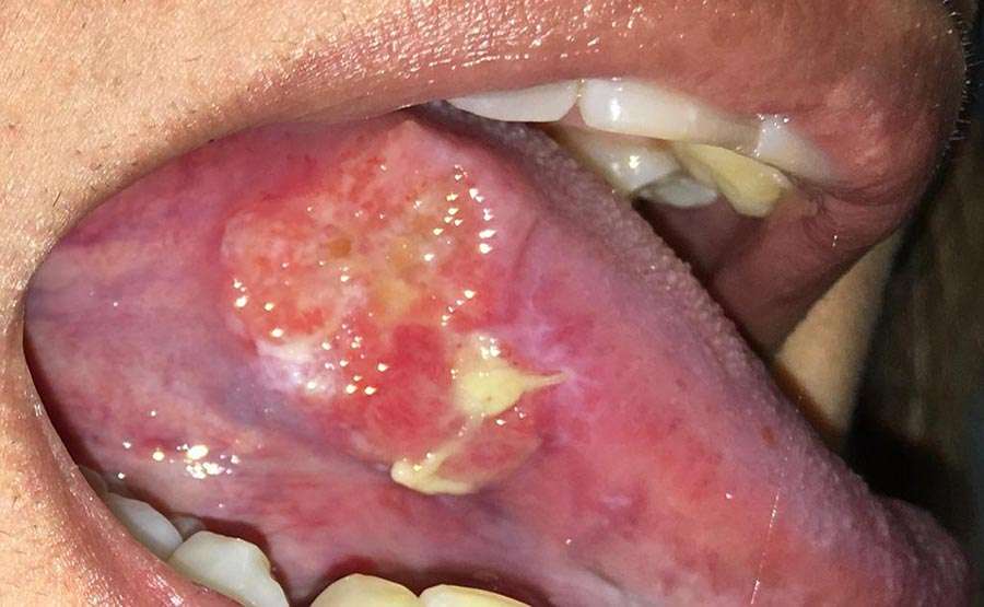
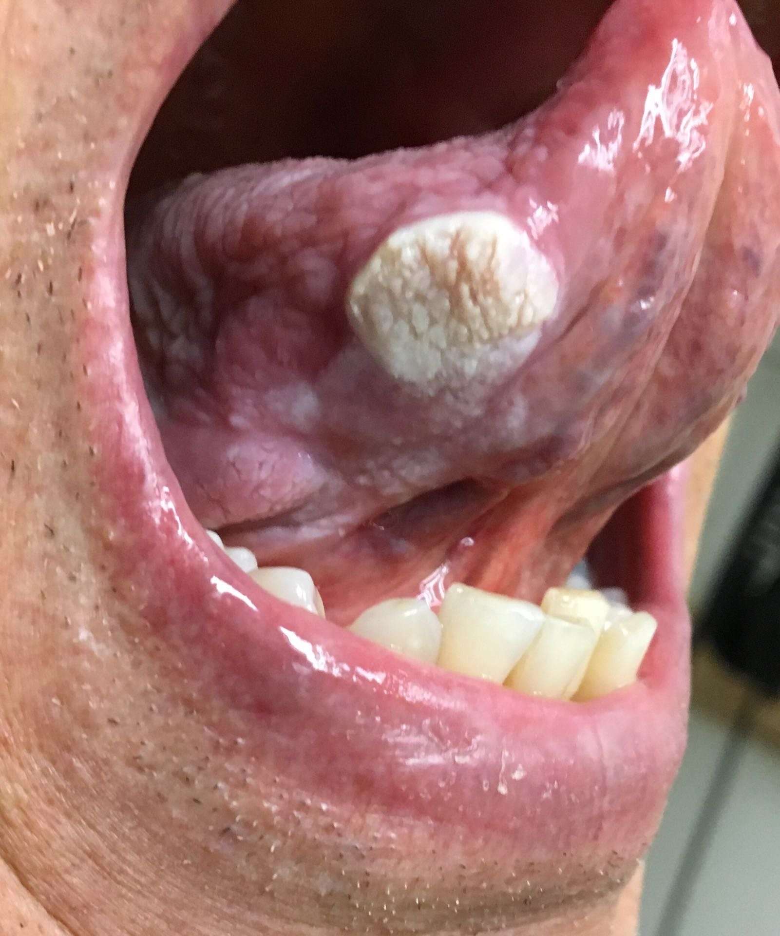
What Are The Signs And Symptoms Of Squamous Cell Carcinoma Of Oral Cavity
The signs and symptoms of Squamous Cell Carcinoma of Oral Cavity include:
- In majority of the cases, the condition is asymptomatic and does not present any signs or symptoms
- Generally, squamous cell carcinomas are slow-growing tumors though SCC of Oral Cavity is an aggressive form of cancer
- The mouth parts affected may include the cheek, hard and soft palate, gums, etc.
- The skin lesions may appear as crusted ulcer, plaques, and nodules
- It may ulcerate and bleed. Occasionally, after the ulcer heals, it may become ulcerated again
- The size of the lesions range from 1-10 cm average size is usually less than 3 cm
- Individuals with immunocompromised states have more aggressive tumors
- Due to the presence of the lesion on the oral mucosa, it may be difficult for the individual to consume food and drink. Also, speaking may be difficult and painful
Asleep In Dentists Chair
It was quite a day. After my transplant, my platelets still had not risen to normal levels. I needed platelets for clotting during the surgery , so, first, I got a platelet infusion.
To stave off the allergic reaction I had had in the past, I took a Benadryl. It made me drowsy. A dentist back home had told me to take two 1-milligram Ativan tablets before surgery instead of getting anesthesia. So I did that in addition to the Benadryl. My sister practically had to drag me to the surgeons office. I could barely keep my mouth open.
How Is Squamous Cell Carcinoma Of Tongue Treated
Early diagnosis and treatment of Squamous Cell Carcinoma of Tongue is important to avoid complications such as metastasis to other regions. The treatment measures may include:
- In most cases, a wide surgical excision and removal of the entire tumor is the preferred treatment option . This may be followed by radiation therapy and/or chemotherapy
- If the tumor has metastasized , then a combination of chemotherapy, radiation therapy, and invasive procedures may be used to treat the tumor
- Targeted therapy medications are generally used for locally infiltrated or metastatic SCCs. This therapy destroys the tumor cells by acting against the proteins that are responsible for tumor growth
- Post-operative care is important: One must maintain minimum activity levels, until the surgical wound heals
- Follow-up care with regular screening and check-ups are important and encouraged
Also Check: Invasive Ductal Carcinoma Grade 2 Survival Rate
My Squamous Cell Carcinoma Of The Tongue
To back up, after my last bone marrow transplant, I have had more squamous cell cancers of the skin than I can count. The most common type of oral cancer is also a squamous cell carcinoma.1 Squamous cells are the flat, skin-like cells that cover the lining of the mouth, nose, larynx, thyroid and throat.2 Squamous cell carcinoma is the name given to cancer that starts in these cells.2
Half Of My Tongue Is Gone But I Couldnt Be Happier

Ive had four of my back teeth removed, the floor of my mouth rebuilt and more than half of my tongue replaced with an arm muscle. But other than a slight lisp and some scars on my neck and forearm, there are no obvious signs that I once had stage IV squamous cell carcinoma, a type of oral cancer.
Recovery from my oral cancer treatment was not fun. It was hard and scary and painful. But it was also worth it.
Thats why I want to share my story both here and through myCancerConnection, MD Andersons one-on-one cancer support community. I want other people facing a similar diagnosis particularly young mothers to see that theres a bright and happy light at the end of that long, dark, uncomfortable tunnel.
My oral cancer symptoms
My first symptom of oral cancer was a small, white patch on the underside of my tongue. It randomly appeared in 2011, and was very sensitive and painful. Since I was only about six weeks away from delivering my second child, I assumed it was one of the million weird things that can go on in your body during pregnancy.
When it didnt go away in a week or two, I mentioned it to my OB-GYN. She said it wasnt pregnancy-related and sent me to a dentist, who said it wasnt a normal mouth sore and sent me to an oral surgeon. The oral surgeon did a biopsy, which came back negative. He said it was a callus and would resolve on its own.
My oral cancer diagnosis
Traveling to MD Anderson for the best oral cancer treatment
My oral cancer treatment
Don’t Miss: Melanoma Bone Cancer Life Expectancy
Endoscopic Procedures: Panendoscopy Narrow Band Imaging Transoral Robotic Surgery And Transoral Laser Microsurgery
In NCUP of levels I, II, III, and VA of the neck, the next step in the traditional algorithm is the panendoscopy or triple endoscopy, including direct laryngoscopy, rigid or flexible bronchoscopy, and rigid or flexible esophagoscopy . Due to the exceedingly low incidence of clinically occult primary in the lung or esophagus with a metastatic node in the neck, many centers have now abandoned the practice of bronchoscopy and esophagoscopy in the search of the unknown primary. Bronchoscopy and esophagoscopy as endoscopic screening for second primaries, not causative of the neck mass, also remains controversial, but has its advocates . On the other hand direct laryngoscopy and careful endoscopy of the nasopharynx is clearly warranted. Examination under anesthesia is far superior to the office examination at identifying a primary tumor, because of relaxation of the pharyngeal musculature and ability to palpate base of tongue, tonsils and nasopharynx. Any firm nodularity, or bleeding on palpation requires biopsy of these suspicious areas .
The literature prior to the era of HPV-related cancer showed increased survival associated with the initial identification of the primary tumor in patients with NCUP . However more recent publications have not been able to show statistically improved survival rates, although other benefits occur with identification of primary tumors, such as precision in planning radiation ports .
Recommended Reading: Squamous Cell Carcinoma Skin Metastasis
How Is Squamous Cell Carcinoma Of Oral Cavity Diagnosed
A diagnosis of Squamous Cell Carcinoma of Oral Cavity is made by:
- Complete physical examination with detailed medical history evaluation
- Examination by a dermatologist using a dermoscopy, a special device to examine the skin
- Woodâs lamp examination: In this procedure, the healthcare provider examines the skin using ultraviolet light. It is performed to examine the change in skin pigmentation
Although the above modalities can be used to make an initial diagnosis, a tissue biopsy of the tumor is necessary to make a definitive diagnosis to begin treatment.
Tissue biopsy:
- A tissue biopsy of the tumor is performed and sent to a laboratory for a pathological examination. A pathologist examines the biopsy under a microscope. After putting together clinical findings, special studies on tissues and with microscope findings, the pathologist arrives at a definitive diagnosis. Examination of the biopsy under a microscope by a pathologist is considered to be gold standard in arriving at a conclusive diagnosis
- Biopsy specimens are studied initially using Hematoxylin and Eosin staining. The pathologist then decides on additional studies depending on the clinical situation
- Sometimes, the pathologist may perform special studies, which may include immunohistochemical stains, molecular testing, and very rarely, electron microscopic studies to assist in the diagnosis
In case of metastatic SCC, the following diagnostic procedures can be used to procure the tissue sample:
Read Also: Invasive Breast Cancer Survival Rate
How Skin Cancer Progresses
All cancer starts in one part of your body. With SCC, it starts in your skin. From there, cancer cells can spread.
How far your cancer has spread is known as its stage. Doctors assign skin cancers a stage number between 0 and 4.
Stage 4 means your cancer has spread beyond your skin. Your doctor might call the cancer advanced or metastatic at this stage. It means your cancer has traveled to one or more of your lymph nodes, and it may have reached your bones or other organs.
The stage of your cancer and where it is located will help your doctor find the right treatment for you. At stage 4 your cancer may not be curable, but it is still treatable.
Does Squamous Cell Carcinoma Spread Quickly
One of the factors that can affect a patients prognosis is whether the malignancy has metastasized . Once squamous cell carcinoma has spread beyond the skin, the five-year survival rate drops to less than 50 percent. Fortunately, its fairly rare for squamous cell carcinoma to metastasize. Plus, when metastasis does occur, the malignancy generally spreads slowly, with the majority of cases being diagnosed before the cancer has spread past the skins upper layer .
When staging squamous cell carcinoma, physicians will take a number of factors into account, one being the degree to which the cancer has already spread throughout the body. For example:
- At Stage 0, squamous cell carcinoma has not spread beyond the epidermis.
- At Stage 1, squamous cell carcinoma has spread deeper into the patients skin but has not entered any lymph nodes or healthy tissues.
- At Stage 2, squamous cell carcinoma still has not metastasized to any lymph nodes or healthy tissues, but displays at least one high-risk feature, which might include spreading into the skins lower layers or the nerves.
- At Stage 3, squamous cell carcinoma has spread into the patients lymph nodes but has not reached any other organs or tissues.
- At Stage 4, which is the most advanced stage, squamous cell carcinoma has spread to at least one distant organ .
Don’t Miss: Well-differentiated Squamous Cell Carcinoma Prognosis
Factors Affecting Squamous Cell Carcinoma Prognosis
There are a handful of factors that can affect a patients prognosis, including:
- Having a weakened immune system
- The location of the tumortumors found on the face, scalp, fingers and toes spread more easily, as do tumors that arise in an open wound
- If the cancer has recurred
- Larger tumors and those that are growing deep in the skin
Oral Squamous Cell Carcinoma
Oral squamous cell carcinoma is the most common oral malignancy and has generally low RAS mutation rates . In OSCC, RALGAP2 was found downregulated in patient tumor samples relative to healthy tissue and low expression inversely correlated with survival . Low mRNA and protein levels of RALGAP1, RALGAP2 and RALGAP correlate with increased activity of RALA and RALB and enhanced invasion of OSCC cell lines. Reintroduction of RALGAP2 is able to dampen migration and invasion in these cell lines, whereas anchorage-independent and -dependent proliferation are not affected. Combined treatment of cells with DNA methyltransferase, histone methyltransferase and histone deacetylase inhibitors can restore RALGAP2 mRNA levels, suggesting altered histone modifications in the RALGAP2 promotor region in combination with DNA methylation as mechanisms downregulating RALGAP2 mRNA expression . Thus, epigenetic mechanisms might at least in part promote OSCC by reducing RALGAP2 expression and consequently enhancing RAL-GTP level.
Crispian Scully, Narendran Andrew. Robinson, in, 2017
Recommended Reading: Stage 4 Basal Cell Carcinoma Life Expectancy
Array Data Analysis And Gene Ontology Analysis
The CEL files from all datasets were imported into the statistical software R 2.4.1 using Bioconductor . The meta-analysis was performed as described . In brief, the Robust Multi-Array Average expression measures were computed after background correction and quantile normalization for each microarray dataset. Then, expression values of the overlapping probesets between U133A and U133 Plus 2.0 arrays were extracted. Probeset-level quantile normalization was performed across all samples to make the effect sizes similar among the four datasets. To visualize the overall expression patterns, we performed Principal Component Analysis after removing the normal group mean vector separately from each of the four datasets. Finally, for every probeset, a mixed effects model was applied to identify differential expression. For gene i in sample k of experiment j,
In the model, the random effect ijis the laboratory effect, and iis the first-order cancer effect, which is our major focus in the identification of cancer-associated genes. After obtaining the estimates and the p-values of the i‘s of each probeset, we corrected the p-values for false discovery rate . We selected genes at the FDR level of 0.01, and with cancer effect size > 1 . Functional analysis of the differentially expressed genes was carried out using the GOstats package in Bioconductor based on the Gene Ontology Consortium database .
External Beam Radiation Therapy Plus Brachytherapy

High rates of local and regional control of tongue base carcinomas can be achieved by using a combination of EBRT and brachytherapy. Harrison and colleagues29 described 68 patients treated with this approach at Memorial Sloan-Kettering. Forty-eight of those patients had tumors classified as T1 or T2. Patients were treated with 50 to 54 Gy of EBRT to the primary tumor site and upper neck followed by a 20- to 30-Gy boost to the primary tumor using an iridium 192 implant. The 5-year actuarial local control rates were 87% for those with T1 tumors, 93% for those with T2 tumors, and 82% for those with T3 tumors. The 5-year actuarial regional control rate was 96%. Complications, including soft tissue ulceration, mandibular osteonecrosis, and bleeding at the time of catheter removal, occurred in 19% of the patients. Similar results for patients with tongue base carcinomas treated with EBRT and brachytherapy were reported from Stanford University30 and from Long Beach Memorial Medical Center.31 With the high control rates that can be achieved with IMRT, altered fractionation schedules, and concurrent chemoradiation therapy, the use of brachytherapy for the management of tongue base carcinoma has declined, because it requires the patient to undergo significant surgery for no clear benefit in results.
Ashish A. Patel, … R. Bryan Bell, in, 2018
Read Also: Carcinoma Cancer Symptoms
What Is Squamous Cell Carcinoma Of The Tongue
Squamous cell carcinoma is one of the most common types of cancer of the tongue. The symptoms vary depending upon the location of the tumor and the stage of the disease. Medical professionals treat this cancer with radiation, chemotherapy, and surgery.
The symptoms of tongue cancer include a sore throat or pain when swallowing, a sore spot in the mouth that doesn’t heal, or a discolored, reddish-pink or gray spot on the tongue that does not heal. Patients might also experience bleeding from the tongue or numbness of the mouth. Rarely, individuals might experience ear pain or changes in the voice. Patients who have advanced cancer might develop liver problems or anemia.
Squamous cell carcinoma of the tongue usually affects people over 50 years old, and men develop the disease at three times the rate of women. About 80% of all people who develop tongue cancer are smokers. Other risk factors include chewing tobacco or smokeless tobacco, heavy alcohol use, and chronic oral irritation from dental cavities or other problems in the mouth.
What Survival Rates Mean
The survival rate is the percentage of people who live for a certain period of time with this cancer. The number is based on research done on large groups of people with the same stage of cancer.
Experts dont know the exact survival numbers for late-stage SCC, because cancer registries dont track statistics for this cancer. However, your doctor may be able to give you an estimate of your prognosis.
When it comes to surviving cancer, everyone is different. Your outcome will depend on the specific treatments you have and how well you respond to them. Talk to your doctor about your outlook and what it means.
Also Check: Treatment For Stage 3 Melanoma
What Is The Prognosis Of Squamous Cell Carcinoma Of Tongue
- In general, Squamous Cell Carcinoma of Tongue is an aggressive form of cancer. If metastasis is observed, then the prognosis is guarded or unpredictable
- Tumors in their early stage with complete excisional treatment typically have good prognosis
- In cases of metastasis, its prognosis depends upon a set of several factors that include:
- Stage of tumor: With lower-stage tumors, when the tumor is confined to site of origin, the prognosis is usually excellent with appropriate therapy. In higher-stage tumors, such as tumors with metastasis, the prognosis is poor
- The surgical respectability of the tumor
- Overall health of the individual: Individuals with overall excellent health have better prognosis compared to those with poor health
- Age of the individual: Older individuals generally have poorer prognosis than younger individuals
- Whether the tumor is occurring for the first time, or is a recurrent tumor. Recurring tumors have a poorer prognosis compared to tumors that do not recur
- Response to treatment: Tumors that respond to treatment have better prognosis compared to tumors that do not respond so well to treatment
How Is Tongue Cancer Diagnosed
To make a diagnosis, the doctor will take a medical history and ask specific questions about symptoms. A patient’s tongue and neck will be examined and a small, long-handled mirror will be used to look down the throat.
Several tests are used to aid in the diagnosis. These tests include:
- X-rays of the mouth and throat, including CT scans .
- PET scans , which use radioactive materials to identify excessive activity in an organ. This may indicate the tumor is growing.
- Tongue cancer usually requires a biopsy, a small sample of tissue that is removed from a tumor to diagnose cancer. After the surgeon removes the tissue, a pathologist will examine the cells under a microscope. There are different methods to obtain a biopsy:
- Fine needle aspiration biopsy. A thin needle is inserted into the tumor mass and a sample is drawn out by suction into a syringe.
- Incisional biopsy. A sample is removed with a scalpel .
- Punch biopsy. A small circular blade removes a round area of tissue.
You May Like: In Situ Cancer Melanoma