Mutation Analysis Of Thecul2 Candidate Tumour Suppressor Gene In Fcrc
To investigate CUL2 as a candidate familial RCC gene we searched for germline mutations by SSCP analysis of all 20 exons encompassing the CUL2 coding region in nine cases , one with multicentric/bilateral CCRCC aged < 50 years, and two with CCRCC aged < 30 years). In the nine samples analysed, four silent polymorphisms were identified: two novel polymorphisms, G1265A in exon 12 and G2617A in the 3UTR , and two known polymorphisms G2057A and G2538) ).
Papillary Renal Cell Carcinoma
Papillary RCCs originate from cells of the proximal convoluted tubule . Papillary RCCs can be divided, based upon clinical characteristics and genetic features, into type 1 and type 2 tumors .
Papillary RCC Type 1
Patients with type 1 tumors have a better prognosis and are diagnosed at an earlier stage compared to type 2 tumors . Several immunohistochemical markers were proposed to distinguish both types of papillary RCC. However, none of the markers are validated to be used in daily practice. Besides rare hereditary forms characterized by germline mutation in the MET gene , in 15-20% of sporadic papillary type 1 cancers somatic mutations in the MET gene can be found . Alterations in the MET gene or variations of chromosome 7 carrying the MET gene are more frequent and were described in 81% of patients with type 1 carcinomas . This suggests an important role of MET in the pathophysiology of type 1 carcinomas, and MET could be a therapeutic target which is currently investigated in clinical trials.
Papillary RCC Type 2
Immune Cells And Expression Of Pd
Current treatment options for ccRCC are surgery and targeted therapy , as well as ICI . Thus, we checked for the presence of the immune checkpoint proteins PD-1 and PD-L1 in the primary tissues. Whereas the expression of PD-1 ranged from 0 to 80% on the immune cells, the expression of PD-L1 was much lower on the tumor cells ranging from 0 to 5%. The presence of cytotoxic T cells, which are CD8-positive, varied substantially. While some tissues contained more than 100 CD8+ cells per high power field , others showed less than ten CD8+ T cells per HPF. Individual cells stained positive for the cytotoxic immune cell marker granzyme B, which could also be NK cells besides CD8+ T cells . IHC staining of ALI PDOs showed positivity for CD8 and granzyme B . Thus, the persistence of cytotoxic immune cells in ALI PDOs suggested that ALI PDOs were suitable models to test the efficacy of ICI targeting the PD-1/PD-L1 axis.
Table 1. Tumor tissue was stained and examined for CD8, granzyme B, PD-1, and PD-L1 positivity to draw conclusions on therapy response.
Figure 4. Immune cells are preserved in the ALI PDOs. ALI PDOs were stained for CD8 and granzyme B to validate the presence of immune cells. Asterisks indicate positive stained cells. Pictures were taken at 20x magnification.
Also Check: Ductal Carcinoma Breast Cancer Survival Rates
Chromophobe Renal Cell Carcinoma
Chromophobe RCC comprises 3% to 5% of all RCCs and is associated with numerous chromosomal monosomies.153,164-169 Patients have an excellent prognosis.169-172 The cytoplasm stains diffusely positive with Hale colloidal iron stain.173
Most aspirates are very cellular, consisting of tumor cell groups and isolated tumor cells that are generally less cohesive than the cells aspirated from clear cell RCC .174-177 The cells of chromophobe RCC may have a koilocytic appearance as a result of their cytoplasmic and nuclear features, resembling koilocytes seen in Pap smears. These consist of very large cells with prominent cell membranes and abundant fluffy cytoplasm, which is granular but not uniformly so. There may be perinuclear clearing of the cytoplasm. Binucleation is common. Although some nuclei can be very round and bland, in general the nuclei of chromophobe carcinoma have markedly irregular outlines, fine chromatin that can be either very light or dark and hyperchromatic, and marked size variation. Prominent nucleoli are uncommon. These nuclear features are distinctly different from those of most other RCCs. Eosinophilic variants have more granular cytoplasm, whereas typical variants have more clear cytoplasm. The cells stain diffusely positive in a cytoplasmic pattern with Hale colloidal iron stain.
Fig. 4.24. Chromophobe renal cell carcinoma.
Fig. 4.25. Chromophobe renal cell carcinoma.
Fig. 4.26. Chromophobe renal cell carcinoma.
The Kidneys And Ureters

The kidneys are two bean shaped organs, each about the same size as a fist. They are near the middle of your back, one on either side of your spine.
The renal pelvis is in the middle of the kidney. Urine collects here and then drains through a tube called the ureter and into the bladder. When you empty your bladder, the urine leaves the body through a tube called the urethra.
Don’t Miss: Skin Cancer Mayo
How Does Clear Cell Sarcoma Form
We know that in CCS, chromosomes break apart and are put back together in the wrong way. This can cause cells to not work right. In CCS, a gene called EWSR1 joins with a region called ATF1 creating a fusion gene called EWSR1/ATF1. This can also happen with the gene CREB1 . Doctors will look for this change in chromosomes to confirm that your cancer is CCS. This gene fusion happens in almost all cases of CCS. So scientists are trying to figure out how this works so they can find and test new treatments.
Neoadjuvant And Adjuvant Therapy
Neoadjuvant therapy is currently under investigation and available inclinical trials.
There is currently no evidence from a recent systematicreview that adjuvant radiation therapyincreases survival .
Similarly, there is currently no evidence from randomisedphase III trials that medical adjuvant therapy offers a survival benefit. The impact on OSof adjuvant tumour vaccination in selected patients undergoing nephrectomy for T3 renalcarcinomas remains unconfirmed . Results fromprior adjuvant trials studying interferon-alpha and interleukin-2 did not show a survival benefit . Asimilar observation was made in an adjuvant trial of girentuximab, a monoclonal antibodyagainst carboanhydrase IX .
At present, there is no OS data supporting the use of adjuvant VEGFR ormTOR inhibitors. Thus far, several RCTs comparing VEGFR-TKI vs. placebo have been published.One of the largest adjuvant trials compared sunitinib vs. sorafenib vs. placebo .Its interim results published in 2015 demonstrated no significant differences in DFS or OSbetween the experimental arms and placebo . The study publishedits updated analysis on a subset of high-risk patients in 2018, which demonstrated 5-yearDFS rates of 47.7%, 49.9%, and 50.0%, respectively for sunitinib, sorafenib, and placebo, and 5-year OS of 75.2%, 80.2%, and 76.5% . The results indicated that adjuvant therapy with sunitinib orsorafenib should not be given .
7.2.5.1.Summary of evidence andrecommendations for adjuvant therapy
|
Summary of evidence |
Don’t Miss: Melanoma Stage 2 Treatment
Immunohistochemistry In Typical Ccrcc
CCRCC tends to express the low molecular weight cytokeratins characteristic of simple epithelia , in particular, the CK expressed most strongly by epithelial cells of the proximal convoluted tubules . Between 94-100% of CCRCCs stain positively with antibodies against CK18, while 14-40% are positive for CK8.
Immunoreactivity for CK7 and CK19 is less common. Coexpression of these CK has been associated with genomic stability, low grade, and favorable prognosis in CCRCC.
Expression of CK7 was observed in all of the recently reported CCRCC variants with smooth muscle stroma, consistent with their low nuclear grades however, CK19 expression was not determined. Most CCRCCs strongly express the intermediate filament vimentin, whereas positive immunostaining for high-molecular-weight cytokeratins is extremely rare.
Overexpression of the epithelial marker EMA/MUC1 is seen in 77-100% of CCRCCs. The proportion of positive cells increases with tumor grade.
Positive staining for the proximal tubular brush border antigens CD10 and RCC marker is reported for 82-94% and 47-85% of CCRCCs, respectively. Relatively few CCRCCs are positive for expression of E-cadherin , CD117/KIT , parvalbumin , or AMACR . Thus, a standard immunoprofile expected for CCRCC is vimentin+ /EMA+ /CD10+ /RCC marker+ /AMACR- /CK7- /CK19- /CD117- /E-cadherin- /parvalbumin-.
In a 2012 study, epithelial adhesion molecule was shown to impart independent prognostic value, particularly in low grade CCRCC.
Radiographic Investigations To Evaluate Rccmetastases
Chest CT is accurate for chest staging . Use of nomograms tocalculate risk of lung metastases have been proposed based on tumour size, clinical stageand presence of systemic symptoms .These are based on large, retrospective datasets, and suggest that chest CT may be omittedin patients with cT1a and cN0, and without systemic symptoms, anaemia or thrombocythemia,due to the low incidence of lung metastases in this group of patients. There is aconsensus that most bone metastases are symptomatic at diagnosis thus, routine bone imagingis not generally indicated . However, bone scan, brain CT, or MRI may be used inthe presence of specific clinical or laboratory signs and symptoms . A recent prospective comparative blinded study involving 92 consecutive mRCC patientstreated with first-line VEGFR-TKI found that whole-body DWI/MRIdetected a statistically significant higher number of bony metastases compared withconventional thoraco-abdomino-pelvic contrast-enhanced CT, with higher number ofmetastases being an independent prognostic factor for progression-free survival and OS.
Also Check: Stage 2 Cancer Symptoms
Bellini Duct And Medullary Carcinomas
Bellini duct carcinomas are aggressive tumors of cells in the collecting duct system , and patients clinically often present with hematuria. Cytogenetic alterations and deletions of chromosomes 1q, 8p, und 13q are described . There is no distinct mutation that characterizes the subtype, but 29% of all patients present with mutations of NF2 and 24% with mutations of SETD2.
Medullary carcinomas are a rare, highly aggressive variant of RCC occurring in patients of younger age and are associated with sickle cell disease or with the heterozygous carriers of the sickle cell allele . There is a high genetic overlap with proximal urothelial cancer . No specific mutations are currently known because of small patient numbers however, in medullary RCC, loss of SMARCB1 or mutations in the ALK gene have been described . A therapeutically targetable genetic mutation is the amplification of the BCR and ABL genes, known from chronic myeloid leukemia, even if BCR/ABL amplifications are only present in a small number of patients with this subtype . Due to the small patient numbers, no general conclusion for Bellini duct and medullary carcinomas can be drawn.
What Is Clear Cell Renal Cell Carcinoma
Clear cell renal cell carcinoma, or ccRCC, is a type of kidney cancer. The kidneys are located on either side of the spine towards the lower back. The kidneys work by cleaning out waste products in the blood. Clear cell renal cell carcinoma is also called conventional renal cell carcinoma.
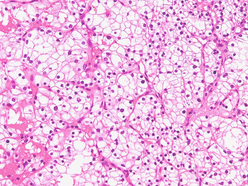
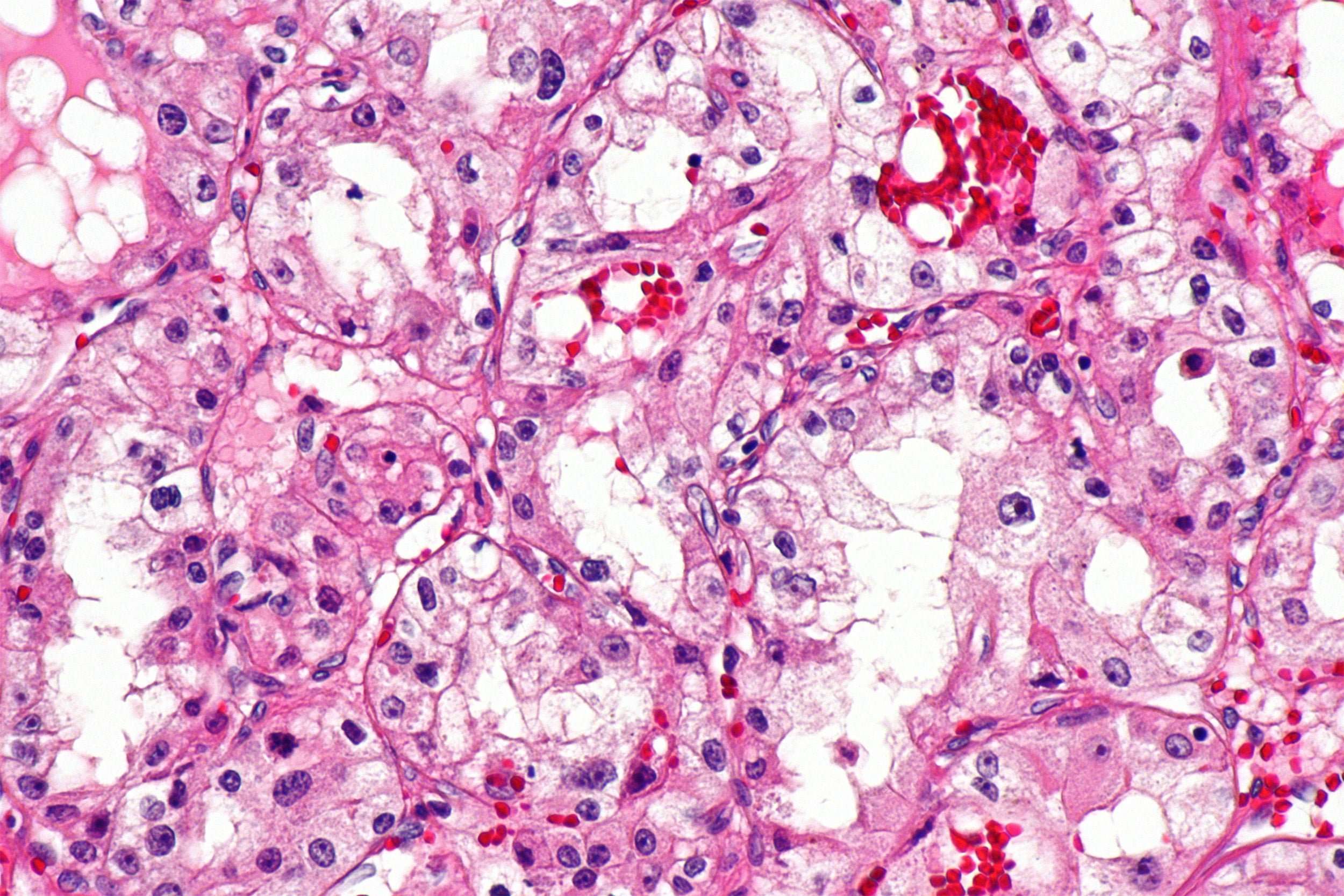
Clear cell renal cell carcinoma is named after how the tumor looks under the microscope. The cells in the tumor look clear, like bubbles.
Read Also: Melanoma 3c
How Will I Feel
The symptoms of kidney cancer are different for each person. In most cases, youâll see blood in your pee. You may feel generally sick, tired, and like you donât want to eat much. And you may have:
- A fever that comes and goes
- A lump in your belly
- Night sweats, so much that you need to change your clothes or sheets
- Pain in your back or side that wonât go away
- Weight loss for no reason
You might also get symptoms where the cancer spreads. If itâs in one of your bones, you might feel pain there. In your lungs, it can give you a cough or trouble breathing.
Overview Of Origin Cell Type Stage And Grade

Human RCCs are thought to arise from a variety of specialized cells located along the length of the nephron. RCC is comprised of several histological cell types. Both clear cell and papillary RCC are thought to arise from the epithelium of the proximal tubule. Chromophobe RCC, oncocytoma, and collecting duct RCC are believed to arise from the distal nephron, probably from the epithelium of the collecting tubule. Each type has differences in genetics, biology and behavior. The most common histological type is clear cell carcinoma, also called conventional RCC, which represents 7580% of RCC. Papillary , chromophobe and other more rare forms such as collecting duct carcinoma comprise the remainder. Oncocytomas represent 37% of renal masses but are invariably benign and their exclusion from classification as RCC has been recommended . Distinct tumors of different cell types can occasionally be seen in the same kidney. An individual tumor can have mixed histologies. The pathologist differentiates cell types routinely by morphology and immunohistochemical markers as well as by cytogenetic and molecular genetic analysis particularly when the cell type is equivocal. Three to five per cent of RCC cannot be classified and are termed RCC, unclassified. Sarcomatoid RCC is no longer considered as a true subtype since sarcomatoid change represents undifferentiated cells associated with progression of disease in all RCC cell types .
Also Check: What Is Braf Melanoma
Kidney Tumor Ali Pdos Resemble Tumor Of Origin Histologically
To verify the similarity between the tumor of origin and our cultivated ALI PDOs, IHC stainings were performed. HE staining revealed that the tumor histology resembled the complex histological structure of the tissue of origin. The growth pattern of the ALI PDOs was solid in most cases . In two cases, the ALI PDOs showed a cystic phenotype.
Figure 2. Kidney tumor ALI PDOs resemble tumor of origin histologically. IHC stainings were prepared to compare the generated ALI PDOs with their derived tissue. ccRCC, pRCC, and urothelial carcinoma show the same tissue structure as their corresponding tissues. Pictures were taken at 10x magnification.
Further IHC analyses showed positivity for PAX8, a marker of renal epithelial origin CA9, a characteristic marker of ccRCC vimentin, a marker for stromal cells and LCA , a marker of lymphocytes . Our results indicate that the complex tissue architecture, phenotype and cellular composition of the primary tumors were maintained.
Figure 3. Immunhistochemistry staining of the cultured ccRCC ALI PDOs. The ALI PDOs show positivity for PAX8, Vimentin, CA9, and LCA.
Recommended Reading: Melanoma Bone Cancer Symptoms
Localtherapy Of Advanced/metastatic Rcc
7.3.1.1.Cytoreductive nephrectomy
Tumour resection is potentially curative only if all tumour deposits areexcised. This includes patients with the primary tumour in place and single- oroligometastatic resectable disease. For most patients with metastatic disease, cytoreductivenephrectomy is palliative and systemic treatments are necessary. In a combined analysisof two RCTs comparing CN+ IFN-based immunotherapy vs. IFN-based immunotherapy only,increased long-term survival was found in patients treated with CN .
The randomised EORTC SURTIME study revealed that the sequence of CN andsunitinib did not affect PFS . The trialaccrued poorly and therefore results are mainly exploratory. However, in secondary endpointanalysis a strong OS benefit was observed in favour of the deferred CN approach in the ITTpopulation with a median OS of 32.4 months in the deferred CN armvs. 15.0 months in the immediate CN arm . The deferred CN approach appears to select out patients with inherent resistanceto systemic therapy . This confirms previous findings fromsingle-arm phase II studies .Moreover, deferred CN and surgery appear safe after sunitinib which supports the findings,with some caution, of the only available RCT.
In patients with poor PS or IMDC poor risk, smallprimaries and high metastatic volume and/or a sarcomatoid tumour, CN is not recommended . These data are confirmed by CARMENA and upfront pre-surgical VEGFR-targeted therapy followed by CNseems to be beneficial .
|
Trial |
|
|---|---|
|
0.68 |
0.57 |
Read Also: Stage 3 Melanoma Survival Rate
What Can I Do
First, work with your doctor to figure out how to best treat it. Even if it canât be cured, you may be able to slow it down and manage your symptoms with surgery, medicine, and other treatments.
You can also do a lot on your own to feel better physically and emotionally:
Pace yourself. Cancer, and even some of its treatments, can wipe you out. Try to keep your days simple and save your energy for the important activities. And donât be shy about resting when you need to.
Speak your symptoms. Your doctor can help with all kinds of common problems from cancer and its treatments, like constipation, upset stomach, and pain. But only if you say something about them. Check in with your doctor often to get the care you need.
Stay active. Exercise lifts your energy and helps you fight off anxiety, depression, and stress. Ask your doctor whatâs safe for you to do.
Tend to your body. Along with regular exercise, try to stick to a healthy diet and get the rest you need. If you donât feel like eating much, a dietitian might be able to help.
Find ways to relax. Itâll keep your mood and energy up. Take time to read a book, go for a walk, call a friend, get a massage, or try some meditation. Or all of the above. Go with works best for you.
Work with your doctor, and try to stay positive. There are more ways to treat the condition than ever before. Your doctor can help you think about which ones are best for you.
Show Sources
Met And Egfr Inhibitors
In addition to VEGF-TKI data on MET inhibition with tivantinib in comparison to a combination of tivantinib with the epidermal growth factor receptor inhibitor erlotinib were recently published . Although an earlier study with erlotinib as monotherapy indicated promising results with an ORR of 11%, the more recent study was stopped permanently after the interim analysis due to a lack of efficacy in both treatment arms. So far, the use of MET inhibitors or erlotinib is not recommended outside clinical trials.
Recommended Reading: Stage Iii Melanoma Treatment
Symptoms Of Metastatic Renal Cell Carcinoma
Your renal cell cancer might not produce symptoms until it spreads outside your kidney. Your first symptoms may be caused by the effects of metastatic cancer in a different part of your body besides your kidney:
- Back pain can occur due to renal cell carcinoma metastasis to the spine
- Breathing problems or feeling faint can occur due to the spread of renal cell carcinoma to the lungs or heart
- Headaches or weakness on one side of the body
- Behavioral changes, confusion, or seizures can occur if renal cell carcinoma spreads to the brain