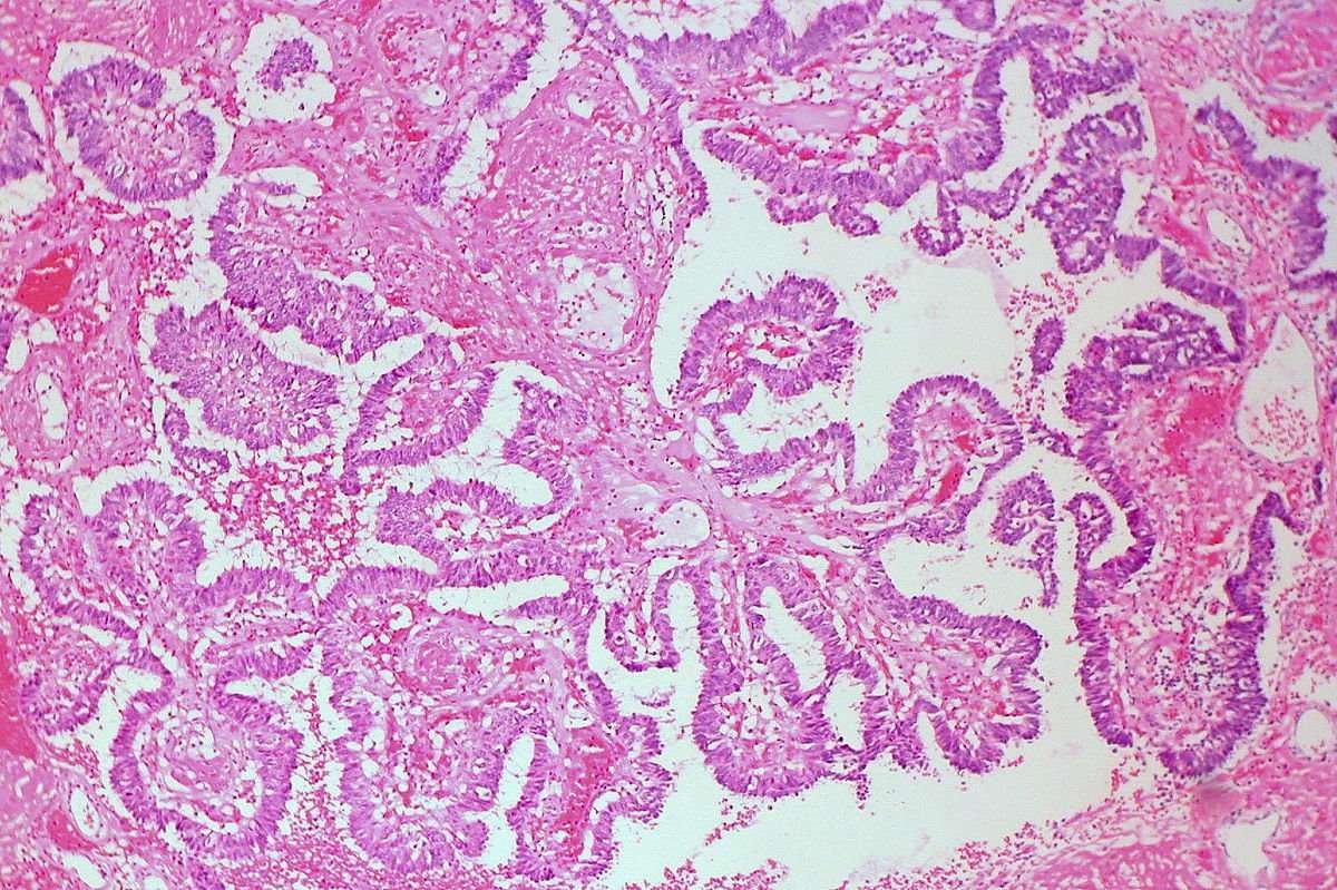
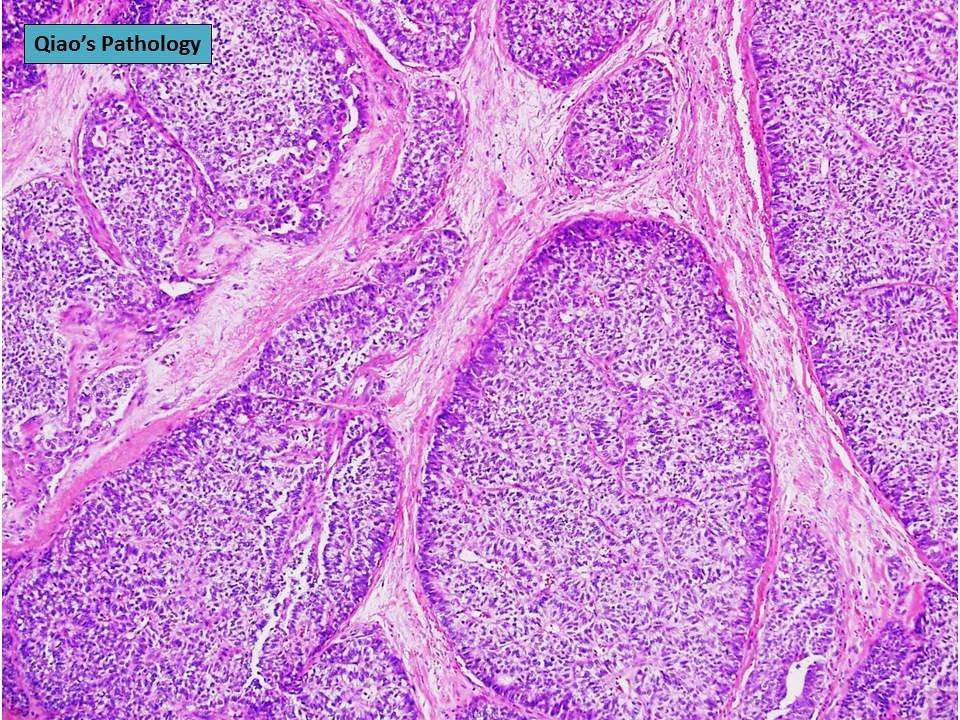
Solid Papillary Carcinoma Of The Breast: A Pathologically And Clinically Distinct Breast Tumor
Jinous Saremian, Marilin Rosa Solid Papillary Carcinoma of the Breast: A Pathologically and Clinically Distinct Breast Tumor. Arch Pathol Lab Med 1 October 2012 136 : 13081311. doi:
Solid papillary carcinomas are tumors morphologically characterized by round, well-defined nodules composed of low-grade ductal cells separated by fibrovascular cores. These tumors are rare and affect predominantly older women. Although they are considered in situ carcinomas, debate and uncertainty still exist regarding their true nature, because immunohistochemistry for myoepithelial cells has shown absence of myoepithelial cell layer along the epithelial-stromal interface of the tumor in many cases. Clinically, these tumors present as a palpable, centrally located mass or as bloody nipple discharge. Pathologically, solid papillary carcinomas exhibit low-grade features, and often the tumors display neuroendocrine and mucinous differentiation. In the majority of cases an associated invasive carcinoma is present, with colloid and neuroendocrine carcinomas being the most common. The pathologic differential diagnosis is broad and ranges from benign to malignant lesions. The treatment for solid papillary carcinomas is surgical excision. When invasive carcinoma is not present, the prognosis is excellent.
Core Biopsy Versus Surgical Excision
There is some controversy regarding the optimal management of papillary lesions diagnosed by core-needle biopsy, with some authors advocating observation for benign disease and others suggesting surgical excision of all lesions in the same setting. A study conducted at St. Vincentâs Comprehensive Cancer Center examined 71 papillary lesions identified by core needle biopsy that were later surgically excised. Of these lesions, 47 were benign at the time of core biopsy. At the time of surgical excision, however, 4 cases revealed malignancy and 13 cases demonstrated atypical features. Of 13 lesions characterized as atypical at the time of core biopsy, surgical excision revealed malignancy in 7 lesions . Overall, it was noted that 38% of cases were up-graded between assessment at core needle biopsy and at the time of surgical excision. Thus for these lesions, surgical excision should be considered, especially if they are clinically or histologically worrisome.
Types Of Papillary Breast Cancer
There are several variations of papillary breast cancer.
Benign papillary lesions
- Intraductal papilloma : A single tumor that grows in the milk ducts near the nipple
- Intraductal papillomatosis: Tumors that grow in the milk ducts near the nipple
Atypical papillary lesions
- Intraductal papilloma with atypical hyperplasia: Abnormal growth of cells
- Papilloma with DCIS: Papilloma with ductal carcinoma in situ, a precancerous condition
Malignant papillary lesions
Noninvasive:
- Papillary ductal carcinoma in situ: Begins in the milk duct of the breast, but hasn’t spread outside the duct
- Encapsulated papillary carcinoma: A rare tumor that is contained in one area
- Solid papillary carcinoma: A rare form with solid nodules, mainly affecting older women
Invasive:
- Invasive papillary carcinoma: A very rare form of ductal carcinoma
- Invasive micropapillary carcinoma: A variant of breast carcinoma with a high chance for regional lymph node involvement
Also Check: Invasive Ductal Carcinoma Survival Rate Stage 3
What Is Invasive Ductal Carcinoma
Breast ducts are the passageways where milk from the milk glands flows to the nipple.
Invasive ductal carcinoma is cancer that happens when abnormal cells growing in the lining of the milk ducts change and invade breast tissue beyond the walls of the duct.
Once that happens, the cancer cells can spread. They can break into the lymph nodes or bloodstream, where they can travel to other organs and areas in the body, resulting in metastatic breast cancer.
What Is Invasive Papillary Carcinoma Of Breast

- Breast cancer is the most common type of cancer diagnosed in women. It is a type of cancer in which certain cells in the breast become abnormal, grow uncontrollably, and form a malignant mass . There are various types of breast cancers which include ductal carcinoma and lobular carcinoma
- Invasive Papillary Carcinoma of Breast is a specific type of invasive ductal carcinoma of breast that initially affects the milk ducts and moves on to involve other parts of the breast. A majority of this cancer type is observed in post-menopausal women
- The signs and symptoms of Invasive Papillary Carcinoma of Breast include lumps in the breast, swelling or skin thickening around the region of the lump, and change in breast profile. Complications from this cancer type include treatment side effects such as nausea, vomiting, and hair loss
- In order to treat Invasive Papillary Breast Carcinoma, the healthcare provider may use a combination of therapies that may include surgery, chemotherapy, radiation therapy and targeted therapy, depending on the stage of the tumor
- The prognosis of Invasive Papillary Carcinoma of Breast is generally much better than invasive ductal carcinomas. An early diagnosis and adequate treatment can significantly improve the outcome
Don’t Miss: Melanoma 3b
What Are The Symptoms Of Invasive Ductal Carcinoma
Like other breast cancers, IDC may present as a lump that you or your doctor can feel on a breast exam. But in many cases, at first, there may be no symptoms, Wright says.
That is why it is important to have screening mammograms to detect breast cancers such as invasive ductal carcinoma. A mammogram may detect a lump that is too small for you to feel, or suspicious calcifications in the breast, either of which will lead to further testing.
According to Wright, the following are possible signs of invasive ductal carcinoma and other breast cancers. If you notice any of these, you should contact your doctor right away for further evaluation:
- Lump in the breast
- Nipple discharge, other than breast milk
- Scaly or flaky skin on the nipple or an ulceration on the skin of the breast or nipple. These can be signs of Pagets disease, a different kind of breast cancer that can occur along with IDC.
- Lumps in the underarm area
- Changes in the appearance of the nipple or breast that are different from your normal monthly changes
What Are The Risk Factors For Invasive Papillary Carcinoma Of Breast
The risk factors for Invasive Papillary Carcinoma of Breast may include:
- Gender: Women have a higher risk for developing the condition than men
- Age: The risk increases for women over the age of 55 years post-menopausal women have the highest risk for Mammary Invasive Papillary Carcinoma
- Postmenopausal hormone therapy: Women taking hormone replacement therapy medications containing both estrogen and progesterone for menopause, have a higher risk of developing breast cancer
- History of breast cancer
- Family history of breast cancer: Women with a mother, sister, or daughter diagnosed with Breast Cancer have a higher risk of developing the condition
- Inherited gene mutations: Mutations in certain genes , can lead to a much higher risk
- Radiation therapy: Receiving radiation therapy to the chest or breast area can also increase the risk
- Obesity: Being overweight or obese increases the risk after menopause
- Alcohol consumption
- Menstrual cycle: Women who got their period before the age of 12 years, and those who reached menopause after age 55 have a higher risk. The longer the duration between menarche and menopause, the greater is the risk. This is due to hormonal influences during the reproductive period on the breast tissue
- Reproductive history: Having the first child after the age of 35, or never having a child
- Physical inactivity: A lack of physical exercise , can increase your risk
- Not breastfeeding the child
Also Check: How Long Until Melanoma Spreads
How Is Papillary Carcinoma Treated
The first step in treating your papillary carcinoma is surgery to remove all tumors. Your doctor might remove just the tumors or your entire breast . You can work with your doctor to find out which option is best for you and discuss any plans for breast reconstruction surgery.
You will have a few weeks to recover from surgery before starting radiation therapy. Radiation therapy uses high-energy X-ray beams to target and destroy cancer cells, while leaving healthy cells unharmed. Not all patients may need radiation therapy.
Following radiation therapy, you might have chemotherapy, depending on specific case and stage of papillary carcinoma. Chemotherapy uses medicines that you take as pills or through an IV to kill cancer cells anywhere in your body.
Papillary carcinoma uses estrogen to help tumors grow. To help keep cancer from coming back, your doctor may have you take hormone therapy for five to ten years after youre finished with cancer treatment. Hormone therapy prevents the cells in your breasts from taking in estrogen and prevents your body from making estrogen. Without estrogen, it is less likely that breast cancer cells will grow.
Causes Of Papillary Carcinoma Of The Breast
Scientists are conducting ongoing research to determine what causes this disease to develop. Currently, they have identified some risk factors for papillary breast cancer.
You may have a higher risk if:
- You are female. Papillary breast cancer can affect men, but women are at greater risk.
- You are post-menopausal: The chances of papillary breast cancer developing is higher in postmenopausal women.
- You are over 60 years old. Papillary breast cancer is more common in older women.
- You are African-American: Research shows that African-Americans are more likely to have papillary breast carcinoma.
Don’t Miss: Body Cancer Symptoms
Intraductal Papilloma With Adh Dcis Or Lobular Neoplasia
These lesions harbor a low nuclear grade atypical epithelial proliferation covering a part of the papilloma. In intraductal papilloma with ADH, this proliferation is limited toâ< â3 mm of extent, whereas in intraductal papilloma with DCIS, it spansââ¥â3 mm. The term âatypical papillomaâ has not been adopted by the recent WHO classification. There are no specific clinical or imaging features. Suspicious microcalcifications may be found on mammography.
Fig. 4
Intraductal papilloma with atypical epithelial proliferation of ductal type in an area ofâ< â3 mm size qualifying for the diagnosis of ADH . The atypical epithelial proliferation lacks CK5 immunoreactivity . Intraductal papilloma with lobular neoplasia characterized by a solid proliferation of monomorphic cells with reduced cell cohesion . The lobular neoplasia is characterized by lack of immunoreactivity for CK5 and e-cadherin
Less frequently, foci of lobular neoplasia may be present within an intraductal papilloma and this should be included in the pathology report . E-cadherin and/or immunohistochemistry for catenin may be helpful to highlight the area of lobular type atypia . Intraductal papilloma with lobular neoplasia diagnosed on CNB or VAB does not require excision if radiological and pathological findings are concordant .
Secretory Breast Carcinoma With Papillary Growth Pattern
Secretory breast carcinoma is exceedingly rare and characterized by a pathognomonic recurrent t translocation, which results in the chimeric fusion gene ETV6-NTRK3 . SBC is mostly observed in post-menopausal women although it can occur at any age and also in males . SBC presents as a slowly growing mass sharing radiological features with benign lesions like papillomas . A predominant papillary pattern may be observed on occasion resulting in a challenging diagnosis on CNB . SBC is composed of a heterogeneous cellular component including cells with amphophilic cytoplasm, apocrine aspect or a âbubbly aspectâ due to abundant intracytoplasmic secretions . Eosinophilic extracellular material positive for PAS, mucicarmine, and Alcian blue is consistently present. SBC usually shows a triple negative phenotype, but low ER expression and an ERâââ/PRâ+âphenotype have been observed. S100 and mammaglobin are usually positive expression of GCDFP-15 has been debated . Pan-TRK immunohistochemistry has been suggested as a useful tool to confirm SBC diagnosis, or may be used for the selection of patients eligible for NTRK inhibitor therapy in the metastatic setting . The clinical course of SBC is indolent compared to IBC-NST however, metastatic cases have recently been described .
Fig. 13
Don’t Miss: Invasive Ductal Carcinoma Breast Cancer Survival Rates
Operative And Pathologic Findings
Intracystic Papillary Carcinoma of the Breast-
Appearance of hypoechoic mass on ultrasound
Subgross appearance of the well circumscribed IPC on hematoxylin-and-eosinstained slide.FIGURE 1D
Intracystic Papillary Carcinoma of the Breast-
Appearance of hypoechoic mass on ultrasound
IPC with fibrosed cyst wall and adjacent low-grade ductal carcinoma in situ at 20x.FIGURE 1E
Intracystic Papillary Carcinoma of the Breast-
Appearance of hypoechoic mass on ultrasound
Displaying low-grade monomorphic nuclei at 40x.
Dr. Mugler: What were the FNA findings?
Dr. Singh: On clinical exam, the mass was 1.5 cm and mobile with irregular contours. The aspirate smears were very cellular, with sheets and papillary configurations of overlapping epithelial cells and many detached single cells , some with nuclear atypia, and many with intact cytoplasm. . There were many macrophages in the background, suggesting a cystic component. Malignancy could not be excluded based on these findings, and an excisional biopsy was recommended.
Dr. Mugler: What were the gross findings of the excisional specimen?
Dr. Singh: A 2.3 X 1.3 X 1.2 cm ovoid piece of breast tissue was received that on serial sections showed a firm discrete, white surface. A cyst cavity was not appreciated.
Dr. Mugler: What were the histologic findings in the resection specimen?
How Is Invasive Papillary Carcinoma Of Breast Treated

Treatment options available for individuals with Invasive Papillary Carcinoma of Breast are dependent upon the following:
- Type of cancer
- The staging of the cancer
- Whether the cancer cells are sensitive to certain particular hormones, and
- Personal preferences
In general, breast cancer stages range from 0 to IV. 0 may indicate a small and non-invasive cancer, while IV indicates that the cancer has spread to other areas of the body. Briefly, as per National Cancer Institute , breast cancer is staged as follows:
- Stage 0 : The abnormal cancer cells are confined to their site of origin
- Stage I: The tumor is 2 centimeters in diameter or less, and has not spread outside the breast
- Stage II: The tumor may be up to 5 centimeters in diameter and may have spread to lymph nodes. Another criteria is that the tumor may be larger than 5 centimeters in diameter, but has not spread to surrounding lymph nodes
- Stage III: The tumor may be more than 5 centimeters in diameter and may have spread to several axillary lymph nodes, or to the lymph nodes near the breastbone. The cancer may also have spread to the breast skin/chest wall, causing ulcer-like sores, or a swelling
- Stage IV: The tumor has spread outside the breast and to other organs, such as the bones, liver, lungs, or brain, regardless of its size
If breast cancer is diagnosed, staging helps determine whether it has spread and which treatment options are best for the patient.
Hormone therapy:
Recommended Reading: Well Differentiated Carcinoma
Data Acquisition And Patient Selection
We used the SEER dataset that was released in April 2015, which included data from 18 population-based registries and covered approximately 28% of U.S. cancer patients. Data for tumour location, grade and histology were recorded according to the International Classification of Diseases for Oncology Version 3 . The inclusion criteria used to identify eligible patients were the following: females aged between 18 and 79, unilateral breast cancer, breast cancer as the first and only cancer diagnosis, diagnosis not obtained from a death certificate or autopsy, only one primary site, pathological confirmation of infiltrating ductal carcinoma, not otherwise specified and papillary carcinoma with invasion , surgical treatment with either mastectomy, breast-conserving surgery or unknown type, known ER and PR statuses, American Joint Committee on Cancer stages IIII and known time of diagnosis from January 1, 2003 to December 31, 2012. Patients diagnosed with breast cancer before 2003 were excluded because the World Health Organization did not recognize IPC as a distinct pathological entity until 2003. In addition, patients who were diagnosed with breast cancer after 2012 were not included because the database was only updated up to December 31, 2012 and we wanted to ensure adequate follow-up time. A total of 233,171 patients were included. Of these patients, 524 were diagnosed with IPC and 232,647 were diagnosed with IDC.
Choose New Hope For Papillary Breast Cancer Alternative Treatment
Our cancer care team knows how distressing it is to receive a breast cancer diagnosis, and learning that you have a rare form of the disease can contribute to your heartbreak. We hope it will be reassuring to know that our medical team here at New Hope Unlimited is committed to the latest research and treatment of papillary breast carcinoma, and is here to support you and your family through treatment and survivorship.
Wear your armor of New Hope. Read about our alternative cancer treatment strategy and how we can help you beat papillary breast cancer.
Also Check: Stage 2 Invasive Ductal Carcinoma Survival Rate
Additional And Relevant Useful Information For Invasive Papillary Carcinoma Of Breast:
- Current studies have shown that aromatase inhibitors, medications that block estrogen hormonal effects in the body, reduce the risk of recurrence of breast cancer. Recent studies have shown that treatment using aromatase inhibitors can be given up to 10 years without affecting the quality of life of women
- Tumors that are negative for estrogen receptor, progesterone receptor, and HER2/neu have worse prognosis. Such tumors are called âtriple-negativeâ tumors
Malignant papillary proliferations of breast can be of 2 types:
- Invasive papillary carcinoma
- Intraductal papillary carcinoma in situ: Majority of malignant papillary tumors of breast are intraductal papillary carcinoma in situ
The following DoveMed website links are useful resources for additional information:
Papillary Breast Cancer Treatment
Local therapy is aimed at preventing the cancer from coming back in the breast. Local therapy includes surgery , and may include radiation.
Systemic therapy is used to prevent the disease from coming back or spreading to another part of the body. This may include endocrine therapy, chemotherapy, and therapy that targets the HER2 protein. Often different types of treatment are used together to achieve the best result.
Your treatment plan will be based on the features of the tumor and the stage of the disease . Your oncology team will recommend a treatment plan based on what is known about papillary breast cancer in general and tailored to your specific disease.
We know that it can be stressful to receive a diagnosis of breast cancer, and learning that you have a rare form of the disease can add to your anxiety. We hope it will be reassuring to know that our team at the Center for Rare Breast Tumors is dedicated to latest research and treatment of papillary breast cancer, and is here to support patients and their families through diagnosis, treatment, and survivorship.
Request an Appointment
Don’t Miss: Invasive Ductal Carcinoma Grade 3 Survival Rate