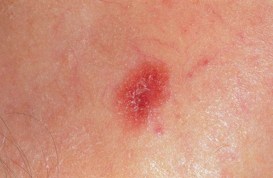
Benign Tumors That Develop From Other Types Of Skin Cells
- Seborrheic keratoses: tan, brown, or black raised spots with a waxy texture
- Hemangiomas: benign blood vessel growths, often called strawberry spots
- Lipomas: soft growths made up of fat cells
- Warts: rough-surfaced growths caused by some types of human papilloma virus
Most of these tumors rarely, if ever, turn into cancers. There are many other kinds of benign skin tumors, but most are not very common.
Plasma And Synchrotron Sources Of Extreme Uv
Lasers have been used to indirectly generate non-coherent extreme;UV radiation at 13.5;nm for . The EUV is not emitted by the laser, but rather by electron transitions in an extremely hot tin or xenon plasma, which is excited by an excimer laser. This technique does not require a synchrotron, yet can produce UV at the edge of the Xray spectrum. can also produce all wavelengths of UV, including those at the boundary of the UV and Xray spectra at 10;nm.
What Does Skin Cancer Look Like
Basal cell carcinoma
-
BCC frequently develops in people who have fair skin. People who have skin of color also get this skin cancer.
-
BCCs often look like a flesh-colored round growth, pearl-like bump, or a pinkish patch of skin.
-
BCCs usually develop after years of frequent sun exposure or indoor tanning.
-
BCCs are common on the head, neck, and arms; however, they can form anywhere on the body, including the chest, abdomen, and legs.
-
Early diagnosis and treatment for BCC are important. BCC can grow deep. Allowed to grow, it can penetrate the nerves and bones, causing damage and disfigurement.
Squamous cell carcinoma of the skin
-
People who have light skin are most likely to develop SCC. This skin cancer also develops in people who have darker skin.
-
SCC often looks like a red firm bump, scaly patch, or a sore that heals and then re-opens.
-
SCC tends to form on skin that gets frequent sun exposure, such as the rim of the ear, face, neck, arms, chest, and back.
-
SCC can grow deep into the skin, causing damage and disfigurement.
-
Early diagnosis and treatment can prevent SCC from growing deep and spreading to other areas of the body.
SCC can develop from a precancerous skin growth
-
People who get AKs usually have fair skin.
-
AKs usually form on the skin that gets lots of sun exposure, such as the head, neck, hands, and forearms.
-
Because an AK can turn into a type of skin cancer, treatment is important.
Melanoma
Also Check: How Can You Get Skin Cancer
Understanding Of Malignant Melanoma Stages
The pathology report can be confusing. It is used by the physician to determine the malignant melanoma stages.
It will list the following:
Type: Is the lesion cutaneous , Mucasol, or Ocular .
The term Breslows Depth of Invasion will be noted which refers to the layers that the cancer has spread down through. Obviously, an increased depth means a far greater chance that it has spread from the site. The cutaneous thickness noted gives you an idea of the survival rate. If the lesion is recorded 4mm deep, then your chance of a five-year survival hovers at 37 to 50 percent. The Clarks Level was once used but has been discarded by pathologists.
Mitotic rate refers to the cells division meaning that if the cells divide rapidly the chance of spreading increases versus a slow-growing tumor.
Additional pathological terms are:
Biological Therapies And Melanoma

Biological therapies are treatments using substances made naturally by the body. Some of these treatments are called immunotherapy because they help the immune system fight the cancer, or they occur naturally as part of the immune system.;There are many biological therapies being researched and trialled, which in the future may help treat people with melanoma. They include monoclonal antibodies and vaccine therapy.;
Don’t Miss: What Are The Symptoms Of Melanoma Skin Cancer
Basal Cell And Squamous Cell Survival Rates
Because basal cell and squamous cell carcinomas are lower-risk skin cancers, theres little information on survival rates based on stage.
Both types of cancer have a very high cure rate. According to the Canadian Cancer Society, the five-year survival rate for basal cell carcinoma is 100 percent. The five-year survival rate for squamous cell carcinoma is 95 percent.
Blockers Absorbers And Windows
Ultraviolet absorbers are molecules used in organic materials to absorb UV radiation to reduce the of a material. The absorbers can themselves degrade over time, so monitoring of absorber levels in weathered materials is necessary.
In , ingredients that absorb UVA/UVB rays, such as , and , are or “blockers”. They are contrasted with inorganic absorbers/”blockers” of UV radiation such as , , and .
For clothing, the represents the ratio of -causing UV without and with the protection of the fabric, similar to ratings for . Standard summer fabrics have UPFs around 6, which means that about 20% of UV will pass through.
Suspended nanoparticles in stained-glass prevent UV rays from causing chemical reactions that change image colors. A set of stained-glass color-reference chips is planned to be used to calibrate the color cameras for the 2019 Mars rover mission, since they will remain unfaded by the high level of UV present at the surface of Mars.
is a deep violet-blue barium-sodium silicate glass with about 9% developed during to block visible light for covert communications. It allows both infrared daylight and ultraviolet night-time communications by being transparent between 320;nm and 400;nm and also the longer infrared and just-barely-visible red wavelengths. Its maximum UV transmission is at 365;nm, one of the wavelengths of .
Read Also: How Do You Get Melanoma Cancer
How Is Melanoma Diagnosed
If you have a mole or other spot that looks suspicious, your doctor may remove it and look at it under the microscope to see if it contains cancer cells. This is called a biopsy.
After your doctor receives the skin biopsy results showing evidence of melanoma cells, the next step is to determine if the melanoma has spread. This is called staging. Once diagnosed, melanoma will be categorized based on several factors, such as how deeply it has spread and its appearance under the microscope. Tumor thickness is the most important characteristic in predicting outcomes.
Melanomas are grouped into the following stages:
- Stage 0 : The melanoma is only in the top layer of skin .
- Stage I: Low-risk primary melanoma with no evidence of spread. This stage is generally curable with surgery.
- Stage II: Features are present that indicate higher risk of recurrence, but there is no evidence of spread.
- Stage III: The melanoma has spread to nearby lymph nodes or nearby skin.
- Stage IV: The melanoma has spread to more distant lymph nodes or skin or has spread to internal organs.
Squamous Cell Carcinoma Causes
Exposure to ultraviolet rays, like the ones from the sun or a tanning bed, affects the cells in the middle and outer layers of your skin and can cause them to make too many cells and not die off as they should. This can lead to out-of-control growth of these cells, which can lead to squamous cell carcinoma.
Other things can contribute to this kind of overgrowth, too, like conditions that affect your immune system.
Don’t Miss: Can Basal Cell Carcinoma Be Fatal
For More Information About Skin Cancer
National Cancer Institute, Cancer Information Service Toll-free: 4-CANCER 422-6237TTY : 332-8615
Skin Cancer Foundation
Media file 1: Skin cancer. Malignant melanoma.
Media file 2: Skin cancer. Basal cell carcinoma.
Media file 3: Skin cancer. Superficial spreading melanoma, left breast. Photo courtesy of Susan M. Swetter, MD, Director of Pigmented Lesion and Cutaneous Melanoma Clinic, Assistant Professor, Department of Dermatology, Stanford University Medical Center, Veterans Affairs Palo Alto Health Care System.
Media file 4: Skin cancer. Melanoma on the sole of the foot. Diagnostic punch biopsy site located at the top. Photo courtesy of Susan M. Swetter, MD, Director of Pigmented Lesion and Cutaneous Melanoma Clinic, Assistant Professor, Department of Dermatology, Stanford University Medical Center, Veterans Affairs Palo Alto Health Care System.
Media file 5: Skin cancer. Melanoma, right lower cheek. Photo courtesy of Susan M. Swetter, MD, Director of Pigmented Lesion and Cutaneous Melanoma Clinic, Assistant Professor, Department of Dermatology, Stanford University Medical Center, Veterans Affairs Palo Alto Health Care System.
Continued
Media file 6: Skin cancer. Large sun-induced squamous cell carcinoma on the forehead and temple. Image courtesy of Dr. Glenn Goldman.
What Tests Are Used To Stage Melanoma
There are several tests your doctor can use to stage your melanoma. Your doctor may use these tests:
- Sentinel Lymph Node Biopsy: Patients with melanomas deeper than 0.8 mm, those who have ulceration under the microscope in tumors of any size or other less common concerning features under the microscope, may need a biopsy of sentinel lymph nodes to determine if the melanoma has spread. Patients diagnosed via a sentinel lymph node biopsy have higher survival rates than those diagnosed with melanoma in lymph nodes via physical exam.
- Computed Tomography scan: A CT scan can show if melanoma is in your internal organs.
- Magnetic Resonance Imaging scan: An MRI scan is used to check for melanoma tumors in the brain or spinal cord.
- Positron Emission Tomography scan: A PET scan can check for melanoma in lymph nodes and other parts of your body distant from the original melanoma skin spot.
- Blood work: Blood tests may be used to measure lactate dehydrogenase before treatment. Other tests include blood chemistry levels and blood cell counts.
Don’t Miss: Is Melanoma The Same As Skin Cancer
What Are The Signs Of Melanoma
Knowing how to spot melanoma is important because early melanomas are highly treatable. Melanoma can appear as moles, scaly patches, open sores or raised bumps.
Use the American Academy of Dermatology’s “ABCDE” memory device to learn the warning signs that a spot on your skin may be melanoma:
- Asymmetry: One half does not match the other half.
- Border: The edges are not smooth.
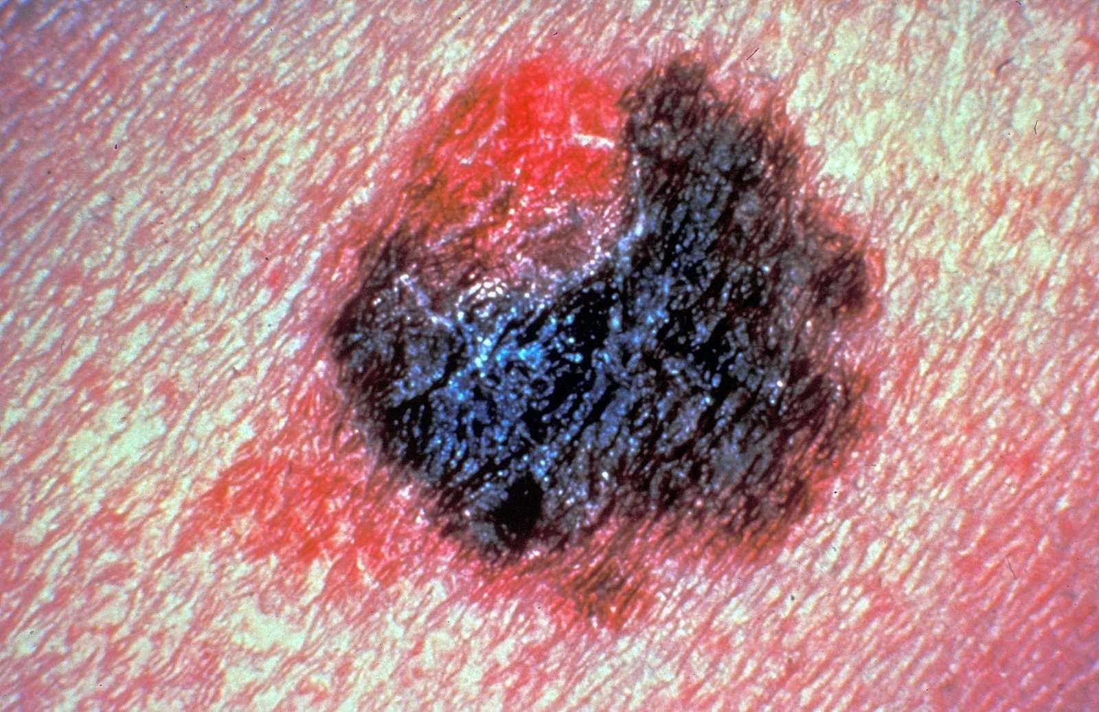
- Color: The color is mottled and uneven, with shades of brown, black, gray, red or white.
- Diameter: The spot is greater than the tip of a pencil eraser .
- Evolving: The spot is new or changing in size, shape or color.
Some melanomas don’t fit the ABCDE rule, so tell your doctor about any sores that won’t go away, unusual bumps or rashes or changes in your skin or in any existing moles.
Another tool to recognize melanoma is the ugly duckling sign. If one of your moles looks different from the others, its the ugly duckling and should be seen by a dermatologist.
Diagnosing Skin Cancer In Dogs

Dog skin cancer is diagnosed by examining the cells of the skin tumor or lesion. Your veterinarian may perform a procedure called a fine needle aspiration, which takes a small sample of cells, or a biopsy, which removes a small portion of the tumor tissue or lesion by surgical incision. These samples are usually sent away to pathology for evaluation in order to obtain an accurate diagnosis.
You May Like: How Can Skin Cancer Kill You
Possible Signs And Symptoms Of Melanoma
The most important warning sign of melanoma is a new spot on the skin or a spot that is changing in size, shape, or color.
Another important sign is a spot that looks different from all of the other spots on your skin .
If you have one of these warning signs, have your skin checked by a doctor.
The ABCDE rule is another guide to the usual signs of melanoma. Be on the lookout and tell your doctor about spots that have any of the following features:
- A is for Asymmetry: One half of a mole or birthmark does not match the other.
- B is for Border:The edges are irregular, ragged, notched, or blurred.
- C is for Color:The color is not the same all over and may include different shades of brown or black, or sometimes with patches of pink, red, white, or blue.
- D is for Diameter:The spot is larger than 6 millimeters across , although melanomas can sometimes be smaller than this.
- E is for Evolving: The mole is changing in size, shape, or color.
Some melanomas dont fit these rules. Its important to tell your doctor about any changes or new spots on the skin, or growths that look different from the rest of your moles.
Other warning signs are:
- A sore that doesnt heal
- Spread of pigment from the border of a spot into surrounding skin
- Redness or a new swelling beyond the border of the mole
- Change in sensation, such as itchiness, tenderness, or pain
- Change in the surface of a mole scaliness, oozing, bleeding, or the appearance of a lump or bump
Exams And Tests For Skin Cancer
If you think a mole or other skin lesion has turned into skin cancer, your primary care provider will probably refer you to a dermatologist. The dermatologist will examine any moles in question and, in many cases, the entire skin surface. Any lesions that are difficult to identify, or are thought to be skin cancer, may then be checked. Tests for skin cancer may include:
- The doctor may use a handheld device called a dermatoscope to scan the lesion. Another handheld device, MelaFind, scans the lesion then a computer program evaluates images of the lesion to indicate if it’s cancerous.
- A sample of skin will be taken so that the suspicious area of skin can be examined under a microscope.
- A biopsy is;done in the dermatologist’s office.
If a biopsy shows that you have malignant melanoma, you may undergo further testing to determine the extent of spread of the disease, if any. This may involve blood tests, a chest X-ray, and other tests as needed. This is only needed if the melanoma is of a certain size.
Continued
Recommended Reading: What Is The Leading Cause Of Skin Cancer
Diagnosis And Treatment Of Malignant Skin Growths
Early diagnosis and treatment of a malignant skin growth are very important.;Complete excision often results in a cure. In fact, complete excision will cure almost all cases of skin cancer if performed in the early stages.
A probable diagnosis of a cancerous skin growth can be madeconsidering some specific factors, including:
- The patients risk factors
- The history of the skin growth and its location
- The appearance of the skin growth
- The texture of the skin growth
A definitive diagnosis can only be made by performing a biopsy and getting the histologic examination results from the lab.
Degradation Of Polymers Pigments And Dyes
is one form of that affects plastics exposed to . The problem appears as discoloration or fading, cracking, loss of strength or disintegration. The effects of attack increase with exposure time and sunlight intensity. The addition of UV absorbers inhibits the effect.
Sensitive polymers include and speciality fibers like . UV absorption leads to chain degradation and loss of strength at sensitive points in the chain structure. Aramid rope must be shielded with a sheath of thermoplastic if it is to retain its strength.
Many and absorb UV and change colour, so and textiles may need extra protection both from sunlight and fluorescent bulbs, two common sources of UV radiation. Window glass absorbs some harmful UV, but valuable artifacts need extra shielding. Many museums place black curtains over and ancient textiles, for example. Since watercolours can have very low pigment levels, they need extra protection from UV. Various forms of , including acrylics , laminates, and coatings, offer different degrees of UV protection.
Don’t Miss: How Bad Is Melanoma Skin Cancer
Squamous Cell Carcinoma Risk Factors
Certain things make you more likely to develop SCC:
- Older age
- Blue, green, or gray eyes
- Blonde or red hair
- Spend time outside, exposed to the sun’s UV Rays
- History of sunburns, precancerous spots on your skin, or skin cancer
- Tanning beds and bulbs
- Long-term exposure to chemicals such as arsenic in the water
- Bowens disease, HPV, HIV, or AIDS
Your doctor may refer you to a dermatologist who specializes in skin conditions. They will:
- Ask about your medical history
- Ask about your history of severe sunburns or indoor tanning
- Ask if you have any pain or other symptoms
- Ask when the spot first appeared
- Give you a physical exam to check the size, shape, color, and texture of the spot
- Look for other spots on your body
- Feel your lymph nodes to make sure they arent bigger or harder than normal
If your doctor thinks a bump looks questionable, theyll remove a sample of the spot to send to a lab for testing.
Continued
Recurrent Basal Cell Carcinoma
Basal cell carcinomas are the most common type of skin cancer, according to the American Cancer Society. These cancers develop within the basal cell layer of the skin, in the lowest part of the epidermis.
Patients who have had basal cell carcinoma once have an increased risk of developing a recurrent;basal cell cancer. Basal cell cancers may recur in the same location that the original cancer was found or elsewhere in the body. As many as 50 percent of cancer patients are estimated to experience basal cell carcinoma recurrence within five years of the first diagnosis.
Basal cell carcinomas typically grow slowly, and it is rare for them to metastasize or spread to nearby lymph nodes or other parts of the body. But early detection and treatment are important.
After completing treatment for basal cell carcinoma, it is important to perform regular self-examinations of the skin to look for new symptoms, such as unusual growths or changes in the size, shape or color of an existing spot. Skin cancers typically develop in areas of the body that are exposed to the sun, but they may also develop in areas with no sun exposure. Tell your oncologist or dermatologist about any new symptoms or suspicious changes you may have noticed.
- Have a history of eczema or dry skin
- Have been exposed to high doses of UV light;
- Had original carcinomas several layers deep in the skin
- Had original carcinomas larger than 2 centimeters
Don’t Miss: Can Merkel Cell Carcinoma Be Cured