Stages Are Used To Describe The Different Types Of Bile Duct Cancer
Intrahepatic bile duct cancer
- Stage 0: In stage 0 intrahepatic bile duct cancer, abnormalcells are found in the innermost layer of tissue lining the intrahepatic bile duct. These abnormal cells may become cancer and spread into nearby normal tissue. Stage 0 is also called carcinoma in situ.
- Enlarge Tumor sizes are often measured in centimeters or inches. Common food items that can be used to show tumor size in cm include: a pea , a peanut , a grape , a walnut , a lime , an egg , a peach , and a grapefruit .
- In stage IA, cancer has formed in an intrahepatic bile duct and the tumor is 5 centimeters or smaller.
- In stage IB, cancer has formed in an intrahepatic bile duct and the tumor is larger than 5 centimeters.
Perihilar bile duct cancer
Symptoms Of Bile Duct Cancer
In most cases, there are no signs of bile duct cancer until it reaches the later stages, when symptoms can include:
- jaundice yellowing of the skin and the whites of the eyes, itchy skin, pale stools and dark-coloured urine
- unintentional weight loss
- abdominal pain
See your GP if you have signs of jaundice or are worried about other symptoms. While it is unlikely you have bile duct cancer, it is best to get it checked.
Read more about preventing bile duct cancer
What Is The Outlook For Extrahepatic Bile Duct Cancer
The only cure for extrahepatic bile duct cancer happens when the cancer can be completely removed.
The outlook for other cases depends on several factors, including overall health, how early the cancer is found and whether it has spread outside of the bile duct .
The five-year survival rate for extrahepatic bile cancer is 10%. This number refers to the number of people who are alive five years after the cancer is found. Remember, though, that these numbers are merely estimates based on reported information.
Recommended Reading: 2nd Stage Cancer
What Is The Intrahepatic Bile Duct
bile ductsIntrahepatic bile ductsbileductshepatic bile ducthepatic bile ductbile
Besides, what does intrahepatic mean?
Medical Definition of intrahepatic: situated or occurring within or originating in the liver intrahepatic cholestasis.
Likewise, what causes dilated intrahepatic ducts? HG Dilated bile ducts are usually caused by an obstruction of the biliary tree, which can be due to stones, tumors , benign strictures , benign stenosis of the papilla , or a
Then, what is intrahepatic bile duct dilation?
Biliary obstruction caused by small simple cysts is very rare. We present a case of biliary dilatation caused by a simple cyst with a 4-cm diameter. Biliary obstruction caused by a simple cyst is very rare,14 and dilatation of the intrahepatic bile duct in association with tumor lesions usually indicates malignancy.
Is a dilated bile duct serious?
Obstruction of any of these bile ducts is referred to as a biliary obstruction. Many of the conditions related to biliary obstructions can be treated successfully. However, if the blockage remains untreated for a long time, it can lead to life-threatening diseases of the liver.
Who Is At Risk

- Chronic infection with hepatitis B virus or hepatitis C virus : Chronicinfection with HBV or HCV increases the risk of liver cancer. HBV and HCV are transmitted through activities that involve percutaneous or mucosal contact with infectious blood or body fluids, including:
- Sex with an infected partner
- Injection drug use that involves sharing needles, syringes, or drug-preparation equipment
- Birth to an infected mother
- Contact with blood or open sores of an infected person
- Needle sticks or sharp instrument exposures
Recommended Reading: Melanoma On Face Prognosis
The Characteristics Of Cca
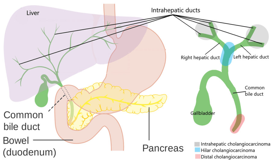
CCA is an epithelial cell malignancy and shows markers of cholangiocytes, which arises from varying locations within the biliary tree. CCA is anatomically classified as intrahepatic, perihilar, and distal CCAs. Intrahepatic CCA is defined as the bile duct from proximal to the second-degree area in the liver. Perihilar CCA is set as the bile duct from the second-degree to the junction of the cystic duct and common bile duct. Distal CCA is defined as the bile duct from the junction to the ampulla of Vater. Intrahepatic CCA is less than 10%, perihilar is 50%, and distal is 40% of cases . Combined hepatocellular CCA accounts for 1% of CCA cases. Intrahepatic CCA is increasing in western countries . The increasing of both the recognition and incidence have led to rising interest in CCAs.
Nataliya Razumilava, … Gregory J. Gores, in, 2018
Facts You Should Know About Bile Duct Cancer
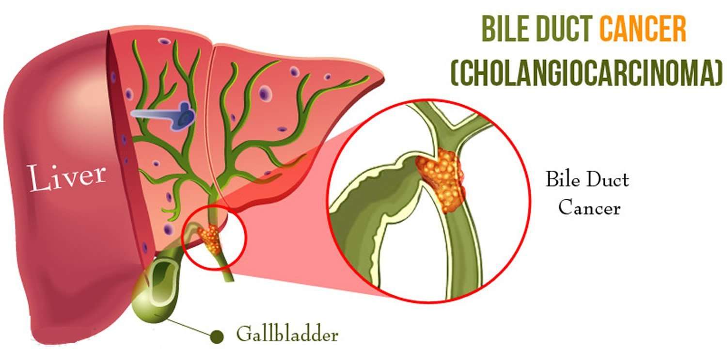
Bile duct cancer arises from the cells that line the bile ducts, the drainage system for bile that is produced by the liver. Bile ducts collect this bile, draining it into the gallbladder and finally into the small intestine where it aids in the digestion process. Bile duct cancer is also called cholangiocarcinoma.
Bile duct cancer is a rare form of cancer, with approximately 2,500 new cases diagnosed in the United States each year. There are three general locations where this type of cancer may arise within the bile drainage system:
- Within the liver affecting the bile ducts located within the liver
- Just outside of the liver located at the notch of the liver where the bile ducts exit
- Far outside of the liver near where the bile ducts enter the intestine
Bile duct cancers are most commonly found just outside of the liver in the perihilar area and least commonly found within the liver.
Also Check: Large Cell Carcinoma Definition
Bile Duct Cancer Is A Rare Disease In Which Malignant Cells Form In The Bile Ducts
A network of tubes, called ducts, connects the liver, gallbladder, and small intestine. This network begins in the liver where many small ducts collect bile . The small ducts come together to form the right and left hepatic ducts, which lead out of the liver. The two ducts join outside the liver and form the common hepatic duct. The cystic duct connects the gallbladder to the common hepatic duct. Bile from the liver passes through the hepatic ducts, common hepatic duct, and cystic duct and is stored in the gallbladder.
When food is being digested, bile stored in the gallbladder is released and passes through the cystic duct to the common bile duct and into the small intestine.
Bile duct cancer is also called cholangiocarcinoma.
There are two types of bile duct cancer:
Intrahepatic Bile Duct Carcinoma
- 2016201720182019202020212022Billable/Specific Code
- C22.1 is a billable/specific ICD-10-CM code that can be used to indicate a diagnosis for reimbursement purposes.
- The 2022 edition of ICD-10-CM C22.1 became effective on October 1, 2021.
- This is the American ICD-10-CM version of C22.1 – other international versions of ICD-10 C22.1 may differ.
type 1 excludes
Also Check: Invasive Lobular Breast Cancer Survival Rate
How Is Cholangiocarcinoma Treated
Your treatment plan for cholangiocarcinoma depends on the location of the cancer and if it has spread. Surgery can treat early bile duct cancers that havent spread. But most bile duct cancers have spread by the time theyre diagnosed. In these cases, your healthcare provider may recommend a combination of multiple treatments.
Bile Duct Cancer Causes And Risk Factors
Experts arenât sure what causes bile duct cancer. Research shows that certain things can raise your chances of getting it, including long-term inflammation from conditions including:
Primary sclerosing cholangitis. This inflammation of your bile duct leads to scarring. Doctors donât know what causes it. Many people who have it also have ulcerative colitis, an inflammation of the large intestine.
Bile duct stones. These are similar to gallstones but much smaller.
Choledochal cysts. Some people are born with a rare condition that causes bile-filled sacs along your bile ducts. Without treatment, they may lead to bile duct cancer.
Liver fluke infection. This is rare in the U.S. but more common in Asia. It happens when people eat raw or poorly cooked fish thatâs infected with tiny parasitic worms called liver flukes. They can live in your bile ducts and cause cancer.
Reflux. When digestive juices from your pancreas flow back into your bile ducts, they canât empty properly.
Cirrhosis. Alcohol and hepatitis can damage your liver and cause scar tissue, raising the risk of bile duct cancer.
Other things that can make you more likely to get bile duct cancer include:
Read Also: Large Cell Carcinoma Lung Cancer
How Is Intrahepatic Bile Duct Cancer Treated
The spread of a patients intrahepatic bile duct cancer, among other factors like age and overall health, will influence how it is treated. Patients with early-stage cancers often have resection surgery to remove the affected bile duct and surrounding lymph nodes. Radiation therapy and chemotherapy may also be used to attack cancer cells. Some later-stage bile duct cancers may not be surgically removable, in which case a combination of radiation therapy, chemotherapy and radiofrequency ablation may be used to help reduce symptoms.
Moffitt Cancer Center offers the latest advances in intrahepatic bile duct cancer treatment within our Gastrointestinal Oncology Program. Our team includes physicians from multiple specialties who focus exclusively on bile duct cancers, giving our patients the best opportunity for a positive outcome and enhanced quality of life. Moffitt also spearheads a groundbreaking clinical trial program that allows eligible patients to receive promising new bile duct cancer therapies, such as immunotherapy, anti-angiogenesis therapies and radiosensitizers.
To consult with a Moffitt oncologist specializing in bile duct cancer about intrahepatic treatment, call or submit a new patient registration form online. We welcome patients with or without referrals.
- BROWSE
How Can I Prevent Extrahepatic Bile Duct Cancer

Since we dont really know what causes extrahepatic bile duct cancer, there is no way to prevent it. However, in general, its a good idea to maintain a healthy lifestyle by eating well and getting enough exercise. If you have other health conditions, like diabetes, stay as healthy as you can. Its wise to avoid excess drinking and avoid drug use, which could help you avoid developing hepatitis and cirrhosis.
You May Like: Well Differentiated
Unblocking The Bile Duct
If your bile duct becomes blocked as a result of cancer, treatment to unblock it may be recommended. This will help resolve symptoms such as:
- jaundice yellowing of the skin and the whites of the eyes
- itchy skin
- abdominal pain
Unblocking the bile duct is sometimes necessary if the flow of bile back into your liver starts to affect the normal functioning of your liver.
The bile duct can be unblocked by using a small tube called a stent. The stent widens the bile duct, which should help to get the bile flowing again.
A stent can be inserted using either:
- a variation of the endoscopic retrograde cholangiopancreatography procedure, which uses an endoscope to guide the stent into the bile duct
- a variation of the percutaneous transhepatic cholangiography procedure, which involves making a small incision in your abdominal wall
Occasionally, an implanted stent can become blocked. If this occurs, it will need to be removed and replaced.
Staging Of Intrahepatic Bile Duct Cancers
After a person is diagnosed with intrahepatic bile duct cancer, doctors will try to figure out if it has spread, and if so, how far. This process is called staging. The stage of a cancer describes how much cancer is in the body. It helps determine how serious the cancer is and how best to treat it. Doctors also use a cancer’s stage when talking about survival statistics.
The earliest stage intrahepatic bile duct cancers are stage 0 . Stages then range from stages I through IV . As a rule, the lower the number, the less the cancer has spread. A higher number, such as stage IV, means cancer has spread more. And within a stage, an earlier letter means a lower stage.
Although each persons cancer experience is unique, cancers with similar stages tend to have a similar outlook and are often treated in much the same way.
Recommended Reading: What Is Stage 2 Squamous Cell Carcinoma
How Does Radiation Therapy Treat Cholangiocarcinoma
Radiation therapy uses powerful beams of radiation to destroy tumors. You might receive radiation therapy after surgery to kill any remaining cancer cells. Or your healthcare provider may suggest it before surgery to shrink tumors before removing them. Radiation can also be delivered through transarterial radioembolization , which uses a catheter to implant tiny beads of radiation in the blood vessels supplying the tumor. The beads block the vessel to prevent blood from getting to the tumor. At the same time, the beads release radiation to shrink the tumor.
What Are The Complications Of Bile Duct Cancer
Obstruction of the bile duct can lead to infection of the bile drainage system or cholangitis.
Cirrhosis may develop in bile duct cancer. This may be due to the tumor obstructing the bile duct and causing liver cell destruction and scarring. This is especially true in patients with primary sclerosing cholangitis. Both cirrhosis and sclerosing cholangitis are listed as potential risk factors for bile duct cancer.
Other complications may be a consequence of the procedures used to diagnose and treat the cancer. These include complications of surgery, chemotherapy, and radiation therapy.
Don’t Miss: Lobular Breast Cancer Survival Rate
Bile Duct & Gallbladder Cancer Risk Factors
Anything that increases your chance of getting biliary cancer is a risk factor. Bile duct and gallbladder cancer risk factors include:
Age: Most cases of biliary cancer in the United States are diagnosed in people between the ages of 50 and 70.
Ethnicity: In the U.S., Native Americans are more likely to get biliary cancers.
Medical conditions: Having any of the following may increase your risk for biliary cancer:
- Primary sclerosing cholangitis : A progressive autoimmune disease which scars the bile ducts over time.
- Chronic liver diseases, including:
Smoking: Smoking increases the risk of many cancers, including bile duct cancer.
Excessive consumption of alcohol: Excessive consumption of alcohol likely increases the risk of biliary cancer. This is especially true for people who have alcohol-associated liver damage.
Family history: Several genetic disorders, including Lynch syndrome, may increase the risk of biliary tract cancers.
Learn more about bile duct cancer:
Treatment Options For Intrahepatic Bile Duct Cancer
The stage of the cancer helps your doctor decide which treatment you need. Treatment also depends on:
- where the cancer is
- your general health and level of fitness
You might have surgery if you have a stage 1 or 2 intrahepatic bile duct cancer. Usually, your surgeon removes part of the liver. This is a major operation. Your doctor will make sure that you are well enough to have it.
Unfortunately, most bile duct cancers are usually advanced by the time they are diagnosed. This means you might not be able to have surgery. Your doctor might suggest other treatments to reduce your symptoms and help you feel better. This includes chemotherapy and putting a small tube to open up a blockage caused by the cancer.
-
AJCC Cancer Staging Manual American Joint Committee on CancerSpringer, 2017.
-
Cancer: Principles and Practice of Oncology VT DeVita, TS Lawrence, SA RosenbergWolters Kluwer, 2019
-
Guidelines for the management and treatment of cholangiocarcinoma: an updateSA Khan and others Gut, 2012. Vol 61, issue 12.
-
Selective internal radiation therapy for unresectable primary intrahepatic cholangiocarcinomaNational Institute for Health and Care Excellence , 2018
-
Biliary cancer: ESMO clinical practice guidelines for diagnosis, treatment and follow upJW Valle and othersAnnals of oncology, 2016. Vol 27, Supplement 5. Pages 28-37
-
Cholangiocarcinoma 2020: the next horizon in mechanisms and managementsJM Banales and others
Also Check: What Does Melanoma In Situ Look Like
Gross Features Of Icc
ICC is grossly classifiable into mass-forming , periductal infiltrating and intraductal growth types. The MF type presents as a nodular lesion or mass in the hepatic parenchyma and the carcinoma is gray to gray-white, firm and solid .1A). The PI type shows spreading of the carcinoma along the portal tracts with stricture of the affected bile ducts and dilatation of the peripheral bile ducts .1B). The IG type presents as a polypoid or papillary tumor within the variably dilated bile duct lumen 1C) and represents the malignant progression of an intraductal papillary neoplasm of the bile duct . ICC arising in the intrahepatic small bile ducts or bile ductules is usually of the MF type while ICC arising in the intrahepatic large bile ducts can be of the PI, MF or IG type. ICC cases involving the hepatic hilum show cholestasis, biliary fibrosis and cholangitis of the intrahepatic bile ducts. MF type ICCs can be quite large. Central necrosis or scarring is common and mucin may be visible on cut surfaces. These three gross types can overlap in a variable combination. At more advanced stages, ICCs consist of variably sized nodules, usually coalescent.
Gross features of intrahepatic cholangiocarcinomas. A: Mass forming type. The carcinoma forms a mass showing compressive growth B: Periductal infiltrating type. The carcinoma spreads along the biliary tree C: Intraductal growth type. The carcinoma shows papillary growth in the dilated intrahepatic bile duct lumen .