Establishment Of A Patient
Neal and colleagues established a protocol to cultivate PDOs in an ALI system, which sustains the complex structure of the tissue of origin . The obtained tissue was fragmented into small pieces and plated within a collagen I matrix on top of a coated insert. Based on this protocol, we established 42 ALI PDOs from different renal tumors, which were surgically resected . Our study included the most common subtypes of RCC, namely ccRCC, and papillary carcinoma . In addition, we successfully cultivated upper urinary tract urothelial carcinomas, which at times occur in the renal pelvis. From the 42 samples, 26 were confirmed as ccRCC . In 77% of the cases, we successfully established ALI PDOs, which could be passaged and remained viable for more than 30 days in culture. The tissue for the cultivated ALI PDOs was obtained from 20 male and six female patients ranging in age from 33 to 87 years. 18 tumors were organ-confined and eight non-organ-confined . The majority of the tumors was graded as G2 , while the remaining tumors were evenly distributed between G1, G3, and G4 .
The success rate for pRCC was 80% and for urothelial carcinoma 88% . In addition, we effectively established ALI PDOs from one oncocytoma , a benign renal tumor. Yet, we failed to cultivate the single case of the rare RCC subtype chromophobe renal cell carcinoma . Overall, cultivation of the ALI PDOs was successful in 72%.
Radiographic Investigations To Evaluate Rccmetastases
Chest CT is accurate for chest staging . Use of nomograms tocalculate risk of lung metastases have been proposed based on tumour size, clinical stageand presence of systemic symptoms .These are based on large, retrospective datasets, and suggest that chest CT may be omittedin patients with cT1a and cN0, and without systemic symptoms, anaemia or thrombocythemia,due to the low incidence of lung metastases in this group of patients. There is aconsensus that most bone metastases are symptomatic at diagnosis thus, routine bone imagingis not generally indicated . However, bone scan, brain CT, or MRI may be used inthe presence of specific clinical or laboratory signs and symptoms . A recent prospective comparative blinded study involving 92 consecutive mRCC patientstreated with first-line VEGFR-TKI found that whole-body DWI/MRIdetected a statistically significant higher number of bony metastases compared withconventional thoraco-abdomino-pelvic contrast-enhanced CT, with higher number ofmetastases being an independent prognostic factor for progression-free survival and OS.
Kidney Tumor Ali Pdos Resemble Tumor Of Origin Histologically
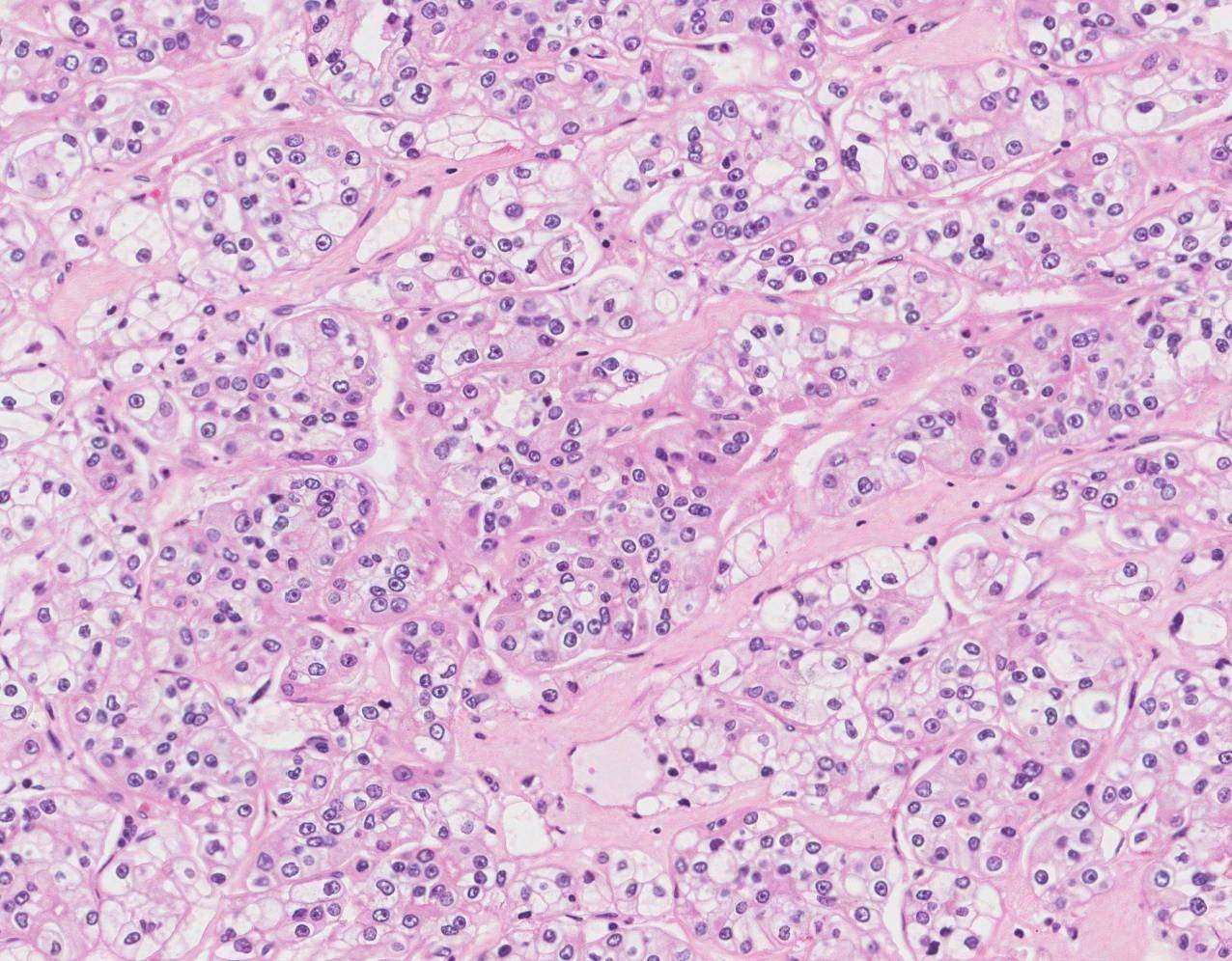
To verify the similarity between the tumor of origin and our cultivated ALI PDOs, IHC stainings were performed. HE staining revealed that the tumor histology resembled the complex histological structure of the tissue of origin. The growth pattern of the ALI PDOs was solid in most cases . In two cases, the ALI PDOs showed a cystic phenotype.
Figure 2. Kidney tumor ALI PDOs resemble tumor of origin histologically. IHC stainings were prepared to compare the generated ALI PDOs with their derived tissue. ccRCC, pRCC, and urothelial carcinoma show the same tissue structure as their corresponding tissues. Pictures were taken at 10x magnification.
Further IHC analyses showed positivity for PAX8, a marker of renal epithelial origin CA9, a characteristic marker of ccRCC vimentin, a marker for stromal cells and LCA , a marker of lymphocytes . Our results indicate that the complex tissue architecture, phenotype and cellular composition of the primary tumors were maintained.
Figure 3. Immunhistochemistry staining of the cultured ccRCC ALI PDOs. The ALI PDOs show positivity for PAX8, Vimentin, CA9, and LCA.
Recommended Reading: Melanoma Bone Cancer Symptoms
Immunohistochemistry In Atypical Ccrcc Variants
Distinguishing between unusual variants of the common RCC histologic types and other types of tumors can be difficult. For example, CCRCCs composed predominantly of granular eosinophilic cells may show a morphological resemblance to the eosinophilic variant of chromophobe RCC or to oncocytoma. Immunohistochemical markers suggested for this distinction include E-cadherin, CD117/KIT, or parvalbumin, which are rarely expressed in CCRCC.
Other markers that may prove useful include the RCC marker and vimentin. These are commonly expressed in CCRCC, including the granular eosinophilic variant. By contrast, the RCC marker is expressed in only 0-4% of chRCC and 0-14% of oncocytomas, while immunostaining for vimentin is positive in only 1-25% of oncocytomas and is negative in chRCC.
Rare tumors composed mainly of clear cells but with a predominant papillary architecture tend to be classified as papillary RCC with extensive clear cell change. However, genetic analysis has, in some cases, found specific translocations characteristic of Xp11 translocation RCC, a tumor with clear and/or eosinophilic cells that can show both papillary architecture and a nested pattern with vascular stroma similar to compact-alveolar CCRCC.
Current And Emerging Therapeutics

The management of metastatic ccRCC has improved dramatically over the last decade . Prior to 2005, only two drugs were approved for the medical treatment of advanced ccRCC and the median survival was poor . The introduction of targeted therapies expanded the treatment options greatly and doubled the median survival . Given the highly vascular nature of ccRCC, tyrosine kinase inhibitors targeting the VEGF signaling pathway provided considerable benefit over the interleukin-2 and interferon treatments. Seven anti-angiogenic drugs have been approved for first-line and second-line treatment of metastatic RCC since 2005, including sorafenib, sunitinib, pazopanib, axitinib, bevacizumab, cabozantinib, and lenvatinib. With the exception of lenvatinib in combination with everolimus and bevacizumab use with interferon-, all approvals for VEGF-targeted therapies have been for single agents. The development of mTOR inhibitors, temsirolimus and everolimus, have provided additional therapeutic benefit as second-line single agents or in the first-line setting in patients with poor risk status. Despite these therapeutic advances, the average duration of disease control with these drugs remains only 8-9 months for first line treatment and 5-6 months in the second line setting. Additional target therapies under investigation also include PT2385, a first-in-class HIF-2 antagonist, which has shown a favorable safety profile and activity in a phase I dose escalation trial.
You May Like: Well Differentiated Squamous Cell Carcinoma Stages
Current And Emerging Therapies For First Line Treatment Of Metastatic Clear Cell Renal Cell Carcinoma
1472274Michael T. Serzan Michael B. Atkins
Georgetown Lombardi Comprehensive Cancer Center, Washington, DC 20057, USA.
Received: Accepted: Available online:Academic Editor: Copy Editor: Production Editor:
© The Author 2021. Open Access This article is licensed under a Creative Commons Attribution 4.0 International License , which permits unrestricted use, sharing, adaptation, distribution and reproduction in any medium or format, for any purpose, even commercially, as long as you give appropriate credit to the original author and the source, provide a link to the Creative Commons license, and indicate if changes were made.
Distinct Cytologic And Tme Features Are Associated With Advanced Tnm Stage And Increased Risk Of Metastasis
Integration of phenotypic subtypes and current prognostic variables. Analysis of the architectural features and disease-free survival corrected for nucleolar grade.p Subgroup analysis of architectural , cytologic and TME features and DFS for each grade. Predictive nomogram in ccRCC patients after nephrectomy.
| Hazard Ratio |
|---|
Read Also: Treatment For Stage 3 Melanoma
Bellini Duct And Medullary Carcinomas
Bellini duct carcinomas are aggressive tumors of cells in the collecting duct system , and patients clinically often present with hematuria. Cytogenetic alterations and deletions of chromosomes 1q, 8p, und 13q are described . There is no distinct mutation that characterizes the subtype, but 29% of all patients present with mutations of NF2 and 24% with mutations of SETD2.
Medullary carcinomas are a rare, highly aggressive variant of RCC occurring in patients of younger age and are associated with sickle cell disease or with the heterozygous carriers of the sickle cell allele . There is a high genetic overlap with proximal urothelial cancer . No specific mutations are currently known because of small patient numbers however, in medullary RCC, loss of SMARCB1 or mutations in the ALK gene have been described . A therapeutically targetable genetic mutation is the amplification of the BCR and ABL genes, known from chronic myeloid leukemia, even if BCR/ABL amplifications are only present in a small number of patients with this subtype . Due to the small patient numbers, no general conclusion for Bellini duct and medullary carcinomas can be drawn.
What Is Clear Cell Renal Cell Carcinoma
Clear cell renal cell carcinoma, or ccRCC, is a type of kidney cancer. The kidneys are located on either side of the spine towards the lower back. The kidneys work by cleaning out waste products in the blood. Clear cell renal cell carcinoma is also called conventional renal cell carcinoma.
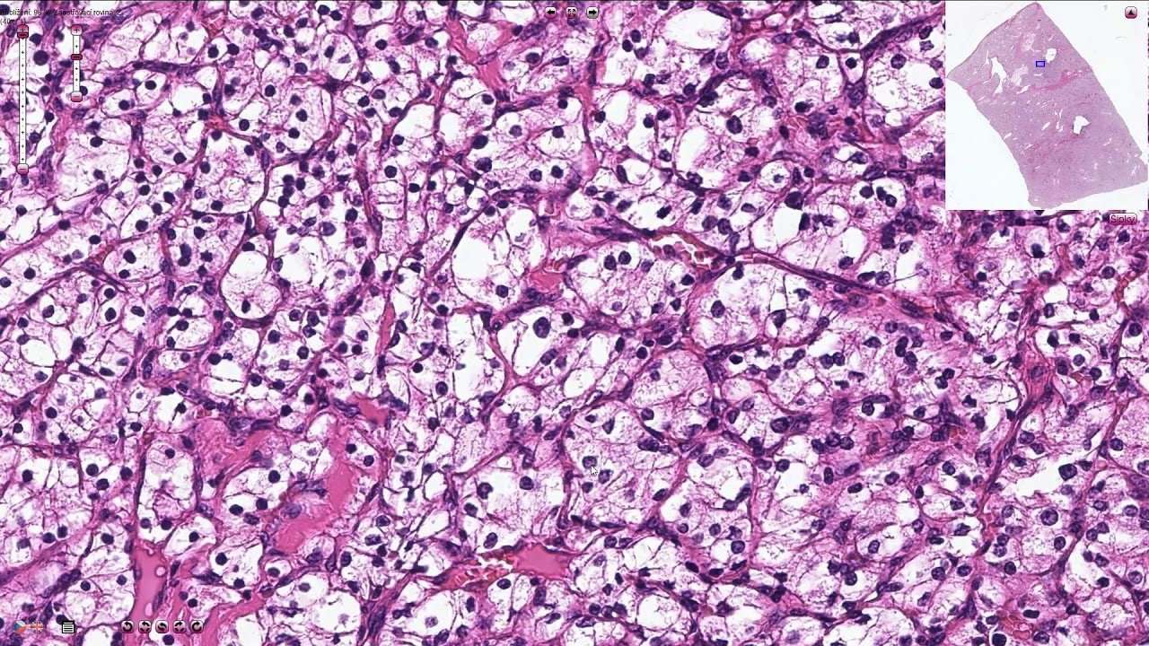
Clear cell renal cell carcinoma is named after how the tumor looks under the microscope. The cells in the tumor look clear, like bubbles.
Read Also: Is Melanoma In Situ Malignant
Localtherapy Of Metastases In Metastatic Rcc
A systematic review of the local treatment of metastases from RCC in anyorgan was undertaken . Interventions included metastasectomy,various radiotherapy modalities, and no local treatment. The outcomes assessed were OS, CSSand PFS, local symptom control and adverse events. A risk-of-bias assessment was conducted. Of the 2,235 studies identified only sixteen non-randomisedcomparative studies were included.
Eight studies reported on local therapies of RCC-metastases in variousorgans . This included metastases to any single organor multiple organs. Three studies reported on local therapies of RCC metastases in bone,including the spine , two in the brain and one each in the liver lung and pancreas . Three studies were published as abstracts only . Datawere too heterogeneous to meta-analyse. There was considerable variation in the type anddistribution of systemic therapies and in reporting theresults.
7.3.2.1.Complete versus no/incompletemetastasectomy
Three studies reported on treatment of RCC metastases inthe lung , liver , and pancreas, respectively. The lung study reported a significantly highermedian OS for metastasectomy vs. medical therapy only for both targeted therapy andimmunotherapy. Similarly, the liver and pancreas study reported a significantly highermedian OS and 5-year OS for metastasectomy vs. no metastasectomy.
7.3.2.2.Local therapies for RCC bonemetastases
7.3.2.3.Local therapies for RCC brainmetastases
7.3.2.4.Embolisation of metastases
7.4.2.2.Interleukin-2
How Is Ccrcc Treated
Treatments for people with ccRCC include surgery and immunotherapy. Treatment will depend on how much the cancer has grown.
Surgery: Once ccRCC is diagnosed, you may have surgery to remove the cancer and part of the kidney surrounding it. In early stage ccRCC, part of the kidney with the cancer is taken out. If ccRCC is in the middle of the kidney, or if the tumor is large, sometimes the entire kidney must be removed. In later stage ccRCC, removal of the kidney is controversial but may be appropriate in some patients.
Immunotherapy: Immunotherapy helps the bodys immune system fight the cancer cells.
Targeted therapy: Targeted therapy targets the changes in cancer cells that help them grow, divide, and spread. Some targeted therapies that are used to treat clear cell renal carcinoma include cabozantinib, axitinib, sunitinib, sorafenib, and pazopanib.
Other treatments can be used that do not involve removing the kidney, such as:
- Radiation therapy, which uses radiation to kill the tumor cells
- Thermal ablation, which uses heat to kill the tumor cells
- Crysosurgery, which uses liquid nitrogen to freeze and kill the tumor cells
Recommended Reading: Lobular Breast Cancer Survival Rate
Genomics And Genetics Of Clear Cell Renal Cell Carcinoma: A Mini
Valerie H. Le James J. Hsieh
Molecular Oncology, Department of Medicine, Siteman Cancer Center, Washington University , St. Louis, MO 63110, USA .
Received:First Decision:Revised:Accepted:Science Editor:Copy Editor:Production Editor:
© The Author 2018. Open Access This article is licensed under a Creative Commons Attribution 4.0 International License , which permits unrestricted use, sharing, adaptation, distribution and reproduction in any medium or format, for any purpose, even commercially, as long as you give appropriate credit to the original author and the source, provide a link to the Creative Commons license, and indicate if changes were made.
Managementof Rcc With Venous Tumour Thrombus

Tumour thrombus formation in RCC patients is a significant adverseprognostic factor. Traditionally, patients with venous tumour thrombus undergo surgery toremove the kidney and tumour thrombus. Aggressive surgical resection is widely accepted asthe default management option for patients with venous tumour thrombus .
7.2.4.1.The evidence base for surgery inpatients with venous tumour thrombus
Data whether patients with venous tumour thrombus should undergo surgery isderived from case series only. In one of the largest published studies a higher level ofthrombus was not associated with increased tumour dissemination to LNs, perinephric fat ordistant metastasis . Therefore, all patients withnon-metastatic disease and venous tumour thrombus, and an acceptable PS, should beconsidered for surgical intervention, irrespective of the extent of tumour thrombus atpresentation. The surgical technique and approach for each case should be selected based onthe extent of tumour thrombus.
7.2.4.2.The evidence base for differentsurgical strategies
A systematic review was undertaken which included only comparative studieson the management of venous tumour thrombus in non-metastatic RCC . Only 5 studies were eligible forfinal inclusion, with a high risk of bias across all studies.
7.2.4.3.Summary of evidence andrecommendations for the management of RCC with venous tumour thrombus
Don’t Miss: Skin Cancer Spreading To Lymph Nodes
How Is Clear Cell Sarcoma Treated
Treatment for each person will be unique. You should go to an expert in sarcoma treatment to decide the best approach for your tumor. You can contact MyPART for help finding experts near you.
Surgery: Surgery to remove the tumor and some healthy tissue around it is the best treatment for CCS. In some cases, an entire arm or leg may need to be amputated. If some cancer cells are left behind, there is a greater chance of the cancer coming back in the same spot. Or, it may spread to a different part of the body.
Radiation therapy: Radiation therapy can be used before or after surgery if doctors think that surgery alone will not remove all the tumor cells. Even so, we dont know if radiation therapy will help you live longer.
Chemotherapy: When surgery is not possible or when the cancer has spread, chemotherapy can be used to treat CCS. But it does not seem to be an effective way to treat CCS.
The Most Frequent Immune Cells In Ccrcc Tumors Are Macrophages Cd4+ T
It has been found in the experimental studies that T cells are the main immune cell population in the ccRCC tumors,. Results of experimental study done by Chevrier et al. show that macrophages are the most frequent immune cells in most ccRCC tumors with a mean of 31% followed by CD8+ T-cells and CD4+ T-cells, respectively , which are in agreement with the results of CIBERSORTx applied on TCGA data set .
You May Like: Invasive Ductal Carcinoma Grade 2 Survival Rate
Monoclonalantibody Against Circulating Vegf
7.4.4.1.Bevacizumab monotherapy andbevacizumab plus IFN-
Bevacizumab is a humanised monoclonal antibody. The double-blind AVORENstudy compared bevacizumab plus IFN- with IFN- monotherapy in mRCC. Overall response was higher in thebevacizumab plus IFN- group. Median PFS increased from5.4 months with IFN- to 10.2 months with bevacizumab plusIFN-. No benefit was seen in MSKCC poor-risk patients.Median OS in this trial, which allowed crossover after progression, was not greater in thebevacizumab/IFN- group .
An open-label trial of bevacizumab plusIFN- vs. IFN- showed ahigher median PFS for the combination group . Objective response rate was also higher in the combinationgroup. Overall toxicity was greater for bevacizumab plus IFN-, with significantly more grade 3 hypertension, anorexia,fatigue, and proteinuria. Bevacizumab, alone, or in combinations, is not widely recommendedor used in mRCC due to more attractive alternatives.
Haematoxylin And Eosin Staining
For haematoxylin and eosin stains the grown ALI PDOs within the collagen gels were cut out with a scalpel and fixed in formalin for 30 min. Subsequently, the fixed ALI PDOs were washed three times with PBS and HistoGel was added according to the manufacturers protocol. The samples were left to cool down and solidify at 4°C and embedded in paraffin. HE staining was performed according to established staining protocols of our routine laboratory.
Recommended Reading: Stage 3b Melanoma Survival Rate
How Does Clear Cell Sarcoma Form
We know that in CCS, chromosomes break apart and are put back together in the wrong way. This can cause cells to not work right. In CCS, a gene called EWSR1 joins with a region called ATF1 creating a fusion gene called EWSR1/ATF1. This can also happen with the gene CREB1 . Doctors will look for this change in chromosomes to confirm that your cancer is CCS. This gene fusion happens in almost all cases of CCS. So scientists are trying to figure out how this works so they can find and test new treatments.
Role Oflymph Node Invasion In Locally Advanced Rcc
In locally advanced RCC, the role of LND is still controversial. The onlyavailable RCT demonstrated no survival benefit for patients undergoing LND but this trialmainly included organ-confined disease cases . In the settingof locally advanced disease, several papers addressed the topic with contradictory results,as did several systematic reviews. Bhindi et al. couldnot confirm any survival benefit in patients at high risk of progression treated with LND. More recently, Luo etal. reported a systematic review and meta-analyses showing a survival benefit inpatients with locally advanced disease treated with LND . Morespecifically, thirteen studies on patients with LND and non-LND were identified and includedin the analysis. In the subgroup of locally advanced RCC , LND showed asignificantly better OS rate in patients who had undergone LND compared to those without LND.
7.2.2.1.Management of clinically negativelymph nodes in locally advanced RCC
In case of cN-, the probability of finding pathologically confirmed LNmetastases ranges between 0 and 25%, depending mainly on primary tumour size and thepresence of distant metastases . In case of clinically negativeLNs at imaging, removal of LNs is justified only if visible or palpable during surgery, at least for staging, prognosis and follow-up implicationsalthough a benefit in terms of cancer control has not yet been demonstrated . Whether to extend the LND also toretroperitoneal areas without cN+ remains controversial .
Don’t Miss: What Is The Prognosis For Skin Cancer
Rna Sequencing Demonstrates Close Molecular Similarity Of Ali Pdos And Tissue Of Origin
Figure 5. RNA sequencing shows close molecular relationship. The close molecular relationship of the samples gene expression profiles was projected onto the first two principal components in a PCA plot . Volcano plot displaying the differentially expressed genes. Genes of interest, which are associated with inflammatory response and IL-2 STAT 5 signaling, are indicated in the up-regulated genes, genes expressed in blood are indicated in down regulated genes . Genes correlated with inflammatory response were up-regulated. NES = normalized enrichment score . Genes correlated with IL-2 STAT5 signaling were up-regulated. NES = normalized enrichment score .