Support For Living With Secondary Breast Cancer
Everyones experience of being diagnosed with secondary breast cancer is different, and people cope in their own way.
For many people, uncertainty can be the hardest part of living with secondary breast cancer.
You may find it helpful to talk to someone else whos had a diagnosis of secondary breast cancer.
- Chat to other people living with secondary breast cancer on our online Forum.
- Meet other women with a secondary diagnosis and get information and support at a Living with Secondary Breast Cancer meet-up.
- Live Chat is a weekly private chat room where you can talk about whatevers on your mind.
You can also call Breast Cancer Nows Helpline free on 0808 800 6000.
Image credit: graphic adapted from: Sersa et al.Electrochemotherapy in treatment of tumours. European Journal of Surgical Oncology. 2008. 34: 232240. Adapted by permission under the Creative Commons Attribution-ShareAlike 3.0 license:creativecommons.org/licenses/by-sa/3.0.
When To See A Doctor
It is always vital to seek medical advice early for a skin change, no matter how small it may appear. Make an appointment with your doctor for a skin exam if you notice:
- Any new changes, lesions, or persistent marks on your skin
- A mole that is asymmetrical, has an irregular border, is multicolored, is large in diameter, is evolving, or has begun to crust or bleed
- An “ugly duckling” mole on the skin
- Any changes to your skin that you are concerned about
Melanoma Signs And Symptoms
Melanoma skin cancer is much more serious than basal cell carcinoma and squamous cell carcinoma. It can spread quickly to other organs and causes the vast majority of skin cancer deaths in the United States. Usually melanomas develop in or around an existing mole.
Appearance
Signs and symptoms of melanoma vary depending on the exact type and may include:
- A flat or slightly raised, discolored patch with irregular borders and possible areas of tan, brown, black, red, blue or white
- A firm bump, often black but occasionally blue, gray, white, brown, tan, red or your usual skin tone
- A flat or slightly raised mottled tan, brown or dark brown discoloration
- A black or brown discoloration, usually under the nails, on the palms or on the soles of the feet
See more pictures and get details about different types of melanoma in our dedicated melanoma section.
You May Like: How Do You Treat Melanoma Skin Cancer
Medical Treatment For Skin Cancer
Surgical removal is the mainstay of therapy for both basal cell and squamous cell carcinomas. For more information, see Surgery.
People who cannot undergo surgery may be treated by external radiation therapy. Radiation therapy is the use of a small beam of radiation targeted at the skin lesion. The radiation kills the abnormal cells and destroys the lesion. Radiation therapy can cause irritation or burning of the surrounding normal skin. It can also cause fatigue. These side effects are temporary. In addition, a topical cream has recently been approved for the treatment of certain low-risk nonmelanoma skin cancers.
In advanced cases, immune therapies, vaccines, or chemotherapy may be used. These treatments are typically offered as clinical trials. Clinical trials are studies of new therapies to see if they can be tolerated and work better than existing therapies.
How Are Moles Evaluated

If you find a mole or spot that has any ABCDE’s of melanoma — or one that’s tender, itching, oozing, scaly, doesn’t heal or has redness or swelling beyond the mole — see a doctor. Your doctor may want to remove a tissue sample from the mole and biopsy it. If found to be cancerous, the entire mole and a rim of normal skin around it will be removed and the wound stitched closed. Additional treatment may be needed.
Read Also: What Is The Cure For Skin Cancer
What Are The Symptoms Of Skin Cancer Of The Head And Neck
Skin cancers usually present as an abnormal growth on the skin. The growth may have the appearance of a wart, crusty spot, ulcer, mole or sore. It may or may not bleed and can be painful. If you have a preexisting mole, any change in the characteristics of this spot – such as a raised or an irregular border, irregular shape, change in color, increase in size, itching or bleeding – are warning signs of melanoma. Sometimes the first sign of melanoma or squamous cell cancer is an enlarged lymph node.
Johns Hopkins Head and Neck Cancer Surgery Specialists
Our head and neck surgeons and speech language pathologists take a proactive approach to cancer treatment. Meet the Johns Hopkins specialists who will work closely with you during your journey.
What Does Melanoma Look Like
Melanoma is a type of cancer that begins in melanocytes . Below are photos of melanoma that formed on the skin. Melanoma can also start in the eye, the intestines, or other areas of the body with pigmented tissues.
Often the first sign of melanoma is a change in the shape, color, size, or feel of an existing mole. However, melanoma may also appear as a new mole. People should tell their doctor if they notice any changes on the skin. The only way to diagnose melanoma is to remove tissue and check it for cancer cells.
Thinking of “ABCDE” can help you remember what to look for:
- Asymmetry: The shape of one half does not match the other half.
- Border that is irregular: The edges are often ragged, notched, or blurred in outline. The pigment may spread into the surrounding skin.
- Color that is uneven: Shades of black, brown, and tan may be present. Areas of white, gray, red, pink, or blue may also be seen.
- Diameter: There is a change in size, usually an increase. Melanomas can be tiny, but most are larger than the size of a pea .
- Evolving: The mole has changed over the past few weeks or months.
Melanomas can vary greatly in how they look. Many show all of the ABCDE features. However, some may show changes or abnormal areas in only one or two of the ABCDE features.
Recommended Reading: Can Basal Cell Carcinoma Be Fatal
What Does Squamous Cell Carcinoma Look Like
Squamous cell skin cancers can vary in appearance, but here, weve provided some examples of how it might appear on your skin.
Squamous cell carcinoma initially appears as a skin-colored or light red nodule, usually with a rough surface. They often resemble warts and sometimes resemble open bruises with raised, crusty edges. The lesions tend to develop slowly and can grow into a large tumor, sometimes with central ulceration.
SCCs can occur on any part of the body, but they are more common on areas of skin exposed to the sun like the scalp, ear or face, so pay attention to these areas.
Squamous cell carcinoma usually develops slowly but can spread to the lymph nodes and other organs if left untreated. If caught early though, it is highly treatable. Early detection strategies are crucial for a successful outcome.
You will notice that all these skin cancer pictures are quite different from one another. Note that not all squamous cell cancers have the same appearance so these photos should serve as a general reference for what they can look like.
Squamous Cell Carcinoma In Situ
This photo contains content that some people may find graphic or disturbing.
DermNet NZ
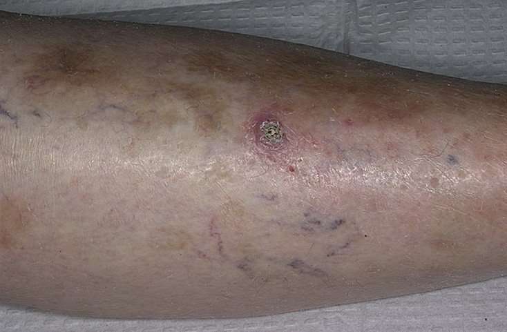
Squamous cell carcinoma in situ, also known as Bowens disease, is a precancerous condition that appears as a red or brownish patch or plaque on the skin that grows slowly over time. The patches are often found on the legs and lower parts of the body, as well as the head and neck. In rare cases, it has been found on the hands and feet, in the genital area, and in the area around the anus.
Bowens disease is uncommon: only 15 out of every 100,000 people will develop this condition every year. The condition typically affects the Caucasian population, but women are more likely to develop Bowens disease than men. The majority of cases are in adults over 60. As with other skin cancers, Bowens disease can develop after long-term exposure to the sun. It can also develop following radiotherapy treatment. Other causes include immune suppression, skin injury, inflammatory skin conditions, and a human papillomavirus infection.
Bowens disease is generally treatable and doesnt develop into squamous cell carcinoma. Up to 16% of cases develop into cancer.
You May Like: How To Recognize Skin Cancer
Getting The Best Treatment
The good news is, weve taken the stress out of seeing a dermatologist. You dont have to look far for excellent dermatology services. Best of all, theres no waiting.
In many parts of New York and throughout the country, patients often wait weeks before they can see a board-certified dermatologist and receive a diagnosis, much less actual treatment.
Thats no longer necessary.
At Walk-in Dermatology, patients can see a board-certified dermatologist seven days a week. Our dermatologists will evaluate your skin and answer all your questions. We will work with you to set up a treatment plan to address your skin condition and get at the root of your issue all convenient to your schedule.
No more waiting days or even weeks to see a dermatologist. Walk-in Dermatology is here for you. We are open and ready to help you regain healthy skin that positively glows with a youthful look.
How Is Psoriasis Treated
Psoriasis is an autoimmune disease. That means it cant be cured. It can, however, be treated to reduce symptoms.
Psoriasis treatments fall into three basic categories. Your doctor may recommend only one of these types of treatments, or they may suggest a combination. The type of treatment you use largely depends on the severity of the psoriasis.
Read Also: How To Self Check For Skin Cancer
What Are The Symptoms Of Skin Cancer
Talk to your doctor if you notice changes in your skin such as a new growth, a sore that doesnt heal, a change in an old growth, or any of the A-B-C-D-Es of melanoma.
A change in your skin is the most common sign of skin cancer. This could be a new growth, a sore that doesnt heal, or a change in a mole.external icon Not all skin cancers look the same.
For melanoma specifically, a simple way to remember the warning signs is to remember the A-B-C-D-Es of melanoma
- A stands for asymmetrical. Does the mole or spot have an irregular shape with two parts that look very different?
- B stands for border. Is the border irregular or jagged?
- C is for color. Is the color uneven?
- D is for diameter. Is the mole or spot larger than the size of a pea?
- E is for evolving. Has the mole or spot changed during the past few weeks or months?
Talk to your doctor if you notice changes in your skin such as a new growth, a sore that doesnt heal, a change in an old growth, or any of the A-B-C-D-Es of melanoma.
How To Spot A Bcc: Five Warning Signs

Check for BCCs where your skin is most exposed to the sun, especially the face, ears, neck, scalp, chest, shoulders and back, but remember that they can occur anywhere on the body. Frequently, two or more of these warning signs are visible in a BCC tumor.
Please note: Since not all BCCs have the same appearance, these images serve as a general reference to what basal cell carcinoma looks like.
An open sore that does not heal
A reddish patch or irritated area
A small pink growth with a slightly raised, rolled edge and a crusted indentation in the center
A shiny bump or nodule
A scar-like area that is flat white, yellow or waxy in color
Recommended Reading: How To Determine Skin Cancer
Basal Cell Carcinoma Early Stages
Basal cells are found within the skin and are responsible for producing new skin cells as old ones degenerate. Basal cell carcinoma starts with the appearance of slightly transparent bumps, but they may also show through other symptoms.
In the beginning, a basal cell carcinoma resembles a small bump, similar to a flesh-colored mole or a pimple. The abnormal growths can also look dark, shiny pink, or scaly red in some cases.
Basal Cell And Squamous Cell Carcinomasigns And Symptoms
The most common warning sign of skin cancer is a change on the skin, especially a new growth or a sore that doesn’t heal. The cancer may start as a small, smooth, shiny, pale or waxy lump. It also may appear as a firm red lump. Sometimes, the lump bleeds or develops a crust.
Both basal and squamous cell cancers are found mainly on areas of the skin that are exposed to the sun the head, face, neck, hands and arms. But skin cancer can occur anywhere.
An early warning sign of skin cancer is the development of an actinic keratosis, a precancerous skin lesion caused by chronic sun exposure. These lesions are typically pink or red in color and rough or scaly to the touch. They occur on sun-exposed areas of the skin such as the face, scalp, ears, backs of hands or forearms.
Actinic keratoses may start as small, red, flat spots but grow larger and become scaly or thick, if untreated. Sometimes they’re easier to feel than to see. There may be multiple lesions next to each other.
Early treatment of actinic keratoses may prevent them from developing into cancer. These precancerous lesions affect more than 10 million Americans. People with one actinic keratosis usually develop more. Up to 1 percent of these lesions can develop into a squamous cell cancer.
Basal cell carcinoma is the most commonly diagnosed skin cancer. In recent years, there has been an upturn in the diagnoses among young women and the rise is blamed on sunbathing and tanning salons.
- Raised, dull-red skin lesion
Read Also: What Is Stage 2 Melanoma Skin Cancer
When Is A Mole A Problem
A mole is a benign growth of melanocytes, cells that gives skin its color. Although very few moles become cancer, abnormal or atypical moles can develop into melanoma over time. “Normal” moles can appear flat or raised or may begin flat and become raised over time. The surface is typically smooth. Moles that may have changed into skin cancer are often irregularly shaped, contain many colors, and are larger than the size of a pencil eraser. Most moles develop in youth or young adulthood. It’s unusual to acquire a mole in the adult years.
Actinic Keratosis Signs And Symptoms
Many people have actinic keratosis , also called solar keratosis, on their skin. It shows that youâve had enough sun to develop skin cancer, and it is considered a precursor of cancer, or a precancerous condition.
Usually AK shows up on the parts of your body that have received the most lifetime sun exposure, like the face, ears, scalp, neck, backs of the hands, forearms, shoulders and lips.
Some of the same treatments used for nonmelanoma skin cancers are used for AK to ensure it does not develop into a cancerous lesion.
Appearance
This abnormality develops slowly. The lesions are usually small, about an eighth of an inch to a quarter of an inch in size. You may see a few at a time. They can disappear and later return.
- AK is a scaly or crusty bump on the skinâs surface and is usually dry and rough. It can be flat. An actinic keratosis is often noticed more by touch than sight.
- It may be the same color as your skin, or it may be light, dark, tan, pink, red or a combination of colors.
- It can itch or produce a prickling or tender sensation.
- These skin abnormalities can become inflamed and be encircled with redness. Rarely, they bleed.
Don’t Miss: What Is The Treatment For Squamous Cell Carcinoma
Tips For Screening Moles For Cancer
Examine your skin on a regular basis. A common location for melanoma in men is on the back, and in women, the lower leg. But check your entire body for moles or suspicious spots once a month. Start at your head and work your way down. Check the “hidden” areas: between fingers and toes, the groin, soles of the feet, the backs of the knees. Check your scalp and neck for moles. Use a handheld mirror or ask a family member to help you look at these areas. Be especially suspicious of a new mole. Take a photo of moles and date it to help you monitor them for change. Pay special attention to moles if you’re a teen, pregnant, or going through menopause, times when your hormones may be surging.
Looking For Signs Of Skin Cancer
Non melanoma skin cancers tend to develop most often on skin that’s exposed to the sun.
To spot skin cancers early it helps to know how your skin normally looks. That way, you’ll notice any changes more easily.
To look at areas you cant see easily, you could try using a hand held mirror and reflect your skin onto another mirror. Or you could get your partner or a friend to look. This is very important if you’re regularly outside in the sun for work or leisure.
You can take a photo of anything that doesn’t look quite right. If you can it’s a good idea to put a ruler or tape measure next to the abnormal area when you take the photo. This gives you a more accurate idea about its size and can help you tell if it’s changing. You can then show these pictures to your doctor.
Also Check: Where Does Skin Cancer Metastasis To