Staging For Basal Cell Carcinoma And Squamous Cell Carcinoma Of The Skin Depends On Where The Cancer Formed
Staging for basal cell carcinoma and squamous cell carcinoma of the eyelid is different from staging for basal cell carcinoma and squamous cell carcinoma found on other areas of the head or neck. There is no staging system for basal cell carcinoma or squamous cell carcinoma that is not found on the head or neck.
Surgery to remove the primary tumor and abnormal lymph nodes is done so that tissue samples can be studied under a microscope. This is called pathologic staging and the findings are used for staging as described below. If staging is done before surgery to remove the tumor, it is called clinical staging. The clinical stage may be different from the pathologic stage.
Wound Care After Mole Removal
Following a mole removal procedure, youll wear a bandage wrapped around the wound for about 24 hours. After 24 hours, youll remove the bandage and clean the wound with gentle, soapy water and a soft gauze or Q-tip.
Its important to rinse the area thoroughly to eliminate leftover soap suds. Dry the skin with another gauze or Q-tip. Apply a thin layer of Vaseline or Aquaphor to the area, and cover the area with a band-aid or bandage. Repeat these steps once a day until you see the wound is healed completely.
You can shower as normal after your mole removal procedure, but dont submerge your bandage or wound in water. Anytime your bandage gets wet, clean the wound thoroughly and replace it with a dry bandage.
Over time, diligent care of your wound will support the skin in healing and recovering fully from the procedure.
What Are The Risks Of Mole Removal
Risks of mole removal methods vary from infection to rare anesthetic allergy and very rare nerve damage. It is always prudent to choose a dermatologist or surgeon with appropriate skills and experience with these removals. This will decrease the risks associated with this procedure.
- Other risks vary depending on the area being treated and the method of removal.
- One of the most common difficulties after mole removal is a scar. Many people will attempt to remove moles for cosmetic reasons, not realizing that each removal will result in a scar. Many times your surgeon can give you an idea of the type of scar after mole removal before you make your decision about removal.
You May Like: Skin Cancer 1st Stage
How Are Tumors Excised From Complex Areas
Excision of tumors on some parts of the body can be tricky. Tricky areas include the face, ear, scalp, sole of the foot, fingers, and toes. If tumors are excised from these areas, it may not be possible to stretch the skin over the site to close it. Special repairs may be needed.1 Examples of repair procedures are skin grafts or flaps. Margins in these areas may be smaller.3
What You Can Expect Before During & After Mole Removal

Like getting your wisdom teeth taken out or having an IUD inserted, mole removal probably isnt high on your cant wait for that appointment! list. How has science not yet invented a way for you to fast-forward to the part where its all over?
Simply thinking about having a mole removed might send a few shivers down your spine, but sometimes its just necessary for your health, Gary Goldenberg, M.D., assistant clinical professor of dermatology at the Icahn School of Medicine at Mount Sinai Hospital, tells SELF. If, for example, you have a mole that your doctor suspects or has confirmed through a biopsy is cancerous, excising the mole can help to stop any cancer from potentially growing more. But people also have moles removed for cosmetic reasons or because theyre simply annoying, like if one falls just under your bra strap and always gets irritated, Dr. Goldenberg says.
No matter the reason youre getting a mole removed, the actual process is pretty much the same for everyone. Heres what you can expect.
For the record, theres technically a difference between having a mole removed and having it biopsied, but these two processes are very closely connected.
Your doctor will typically perform a skin biopsy by using a tool similar to a razor to shave off the mole, using a circular device to remove a section of the mole, or using a scalpel to remove the whole thing, the Mayo Clinic says.
Also Check: Malignant Breast Cancer Survival Rate
How Is Basal Cell Skin Cancer Treated When It Grows Deep Or Spreads
While this skin cancer tends to grow slowly, early treatment is recommended. Without treatment, BCC can grow deep, destroying what lies in its way. This can be disfiguring. The medical term for this is advanced basal cell carcinoma.
Its also possible for BCC to spread to other parts of your body, but this is rare. When the cancer spreads, it typically travels first to the lymph nodes closest to the tumor. From there, it tends to spread through the blood to bones, the lungs, and other parts of the skin. When this skin cancer spreads, it is called metastatic basal cell carcinoma.
For cancer that has grown deep or spread to the closest lymph nodes, treatment may involve:
-
Surgery to remove the tumor
-
Follow-up treatment with radiation to kill any remaining cancer cells
For some patients, medication that works throughout the body may be an option. Medication may also be used to treat cancer that:
-
Returns after surgery or radiation treatments
-
Has spread to another part of the body
Two such medications have been approved by the U.S. Food and Drug Administration . Both come in pill form and are taken every day. A patient only stops taking the medication if the cancer starts to grow, or the side effects become too severe.
The two medications are:
-
Sonidegib
-
Vismodegib
In clinical trials, these medications have been shown to stop or slow down the spread of the cancer and shrink the cancerous tumors in some patients.
Also Check: Large Cell Carcinoma Definition
What Is The Likely Outcome For Someone Who Has Bcc
When found early and treated, this skin cancer can often be removed. However, this skin cancer can return. You also have a higher risk of developing another BCC or other type of skin cancer.
Thats why self-care becomes so important after treatment for BCC. Youll find the self-care that dermatologists recommend at, Basal cell carcinoma: Self-care.
ImageGetty Images
ReferencesBichakjian CK, Armstrong A, et al. Guidelines of care for the management of basal cell carcinoma. J Am Acad Dermatol 2018 78:540-59.
Bichakjian CK, Olencki T, et al. Basal cell skin cancer, Version 1.2016, NCCN Clinical Practice Guidelines in Oncology. J Natl Compr Canc Netw. 2016 14:574-97.
Cameron MC, Lee E, et al. Basal cell carcinoma: Epidemiology pathophysiology clinical and histological subtypes and disease associations. J Am Acad Dermatol 2019 80:303-17.
Cameron MC, Lee E, et al. Basal cell carcinoma: Contemporary approaches to diagnosis, treatment, and prevention. J Am Acad Dermatol 2019 80:321-39.
Nouri K, Ballard CJ, et al. Basal cell carcinoma. In: Nouri K, et al. Skin Cancer. McGraw Hill Medical, China, 2008: 61-81.
Xie P, Lefrançois P. Efficacy, safety, and comparison of sonic hedgehog inhibitors in basal cell carcinomas: A systematic review and meta-analysis. J Am Acad Dermatol 2018 79:1089-100.
Read Also: Lobular Breast Cancer Stage 3
Read Also: Invasive Ductal Carcinoma Survival
Surgery To Remove Melanoma That Has Spread
You might have surgery to remove melanoma that has spread to other areas of the skin or body, such as the lungs, skin and bowel. Cancer that has spread to another area of the body is called secondaries or metastases. The operation you have depends on which part of the body the melanoma has spread to.
For example, you might have surgery to remove a secondary melanoma in the skin. Or it might be possible for some people to have an operation to remove a secondary melanoma in their lung or bowel. This operation is more likely if there are no other signs of melanoma elsewhere in the body. And you need to be reasonably fit and well to have this operation.
It is not usually possible to cure the melanoma. But some people can stay well for months or sometimes years after having several different treatments such as surgery to remove metastases, targeted cancer drugs or immunotherapies.
Simple Excision And Repair
In most cases, melanoma is cut out by simple excision.
- A local anaesthetic injection is given to numb the skin that is to be removed.
- The doctor will cut around and under the melanoma with a scalpel. As described above, a margin of normal skin tissue surrounding the melanoma will also be cut out.
- There might be some bleeding in the area, and the doctor may use a tool to burn and seal off the wound .
- The edges of the wound are sewn together to make a thin line of stitches .
- A dressing will be applied.
- You will be told how to care for your wound and when to get the stitches out.
Don’t Miss: What Is The Survival Rate For Invasive Ductal Carcinoma
What Is The Difference Between A Biopsy And A Removal
A mole biopsy involves one of our doctors taking a small sample of a moles cells by gently shaving the surface of the mole and the surrounding skin with a sharp scalpel. This procedure is performed in our office with local anesthetic and takes just a few minutes. The resulting sample is examined in order to determine whether the cells are normal or show signs of cancer.
Removal can be performed for aesthetic or medical reasons, or a combination of both. Our doctor works carefully to ensure that all of the moles cells are removed. If cells are left over after treatment, a mole can regrow or, even more seriously, cancerous cells from a mole can spread to other areas. Youll return to our office for post-removal check-ups in order for our doctors to check your progress, and we always recommend that our patients have at least one general appointment per year to keep their skin healthy and monitor their moles.
What Are The Signs Of Melanoma
Knowing how to spot melanoma is important because early melanomas are highly treatable. Melanoma can appear as moles, scaly patches, open sores or raised bumps.
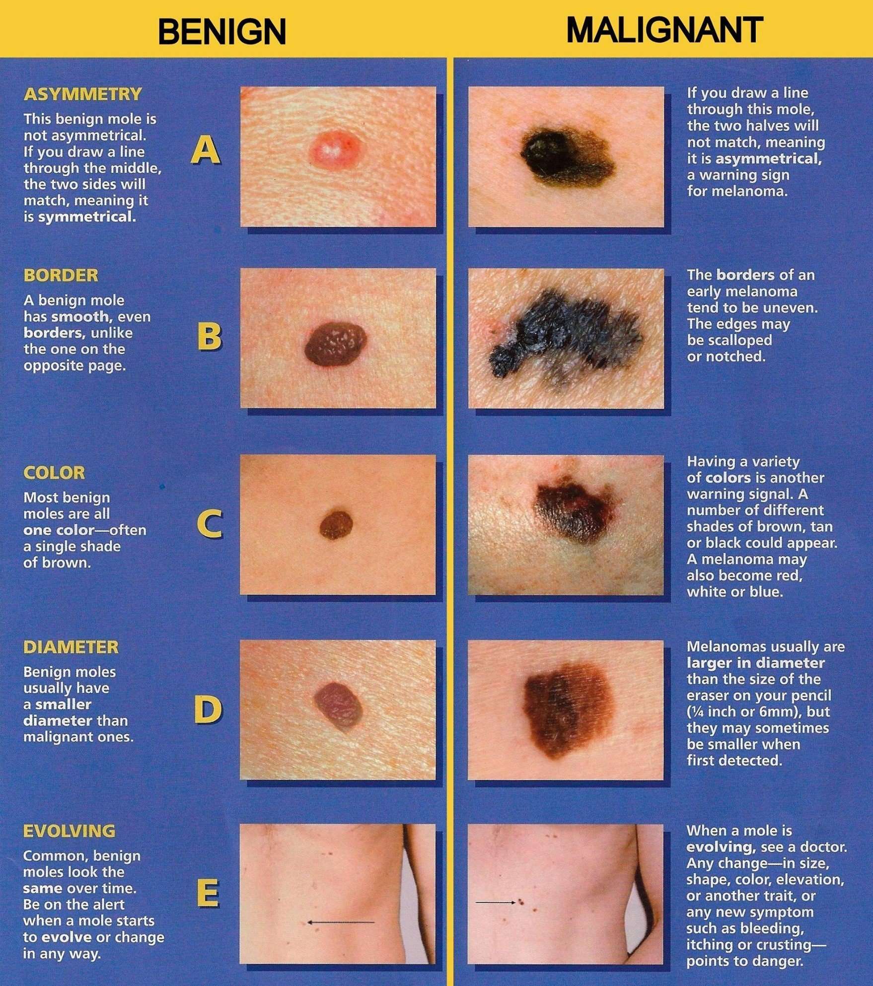
Use the American Academy of Dermatology’s “ABCDE” memory device to learn the warning signs that a spot on your skin may be melanoma:
- Asymmetry: One half does not match the other half.
- Border: The edges are not smooth.
- Color: The color is mottled and uneven, with shades of brown, black, gray, red or white.
- Diameter: The spot is greater than the tip of a pencil eraser .
- Evolving: The spot is new or changing in size, shape or color.
Some melanomas don’t fit the ABCDE rule, so tell your doctor about any sores that won’t go away, unusual bumps or rashes or changes in your skin or in any existing moles.
Another tool to recognize melanoma is the ugly duckling sign. If one of your moles looks different from the others, its the ugly duckling and should be seen by a dermatologist.
Don’t Miss: Well Differentiated Squamous Cell Carcinoma Prognosis
What Does Melanoma Look Like
Often the first sign of melanoma is a change in the shape, color, size, or feel of an existing mole. Melanoma may also appear as a new colored area on the skin.
The “ABCDE” rule describes the features of early melanoma :
- Asymmetry. The shape of one half does not match the other half.
- Border that is irregular. The edges are often ragged, notched, or blurred in outline. The pigment may spread into the surrounding skin.
- Color that is uneven. Shades of black, brown, and tan may be present. Areas of white, gray, red, pink, or blue may also be seen.
- Diameter. There is a change in size, usually an increase. Melanomas can be tiny, but most are larger than 6 millimeters wide .
- Evolving. The mole has changed over the past few weeks or months.
Melanomas can vary greatly in how they look. Many show all of the ABCDE features. However, some may show only one or two of the ABCDE features . Several photos of melanomas are shown here. More photos are on the What Does Melanoma Look Like? page.
Melanoma Photos
In advanced melanoma, the texture of the mole may change. The skin on the surface may break down and look scraped. It may become hard or lumpy. The surface may ooze or bleed. Sometimes the melanoma is itchy, tender, or painful.
How Do Doctors Remove Moles

There are four primary methods of mole removal that doctors or other medical professionals may use. The most common is surgery, in which the mole must be dug out of the skin with a scalpel or sharp knife. Small incisions will usually heal on their own, but larger cuts typically those requiring stitches can sometimes leave a mark or scar. Patients concerned about scarring often look into the second removal options, laser excision or radiosurgery, both of which are less invasive but may not be appropriate for deep moles. Freezing and burning are also possibilities.
The technique that a health care provider chooses to use when removing a mole usually depends on the type of mole at issue. Moles are skin aberrations that are known scientifically as nevi, and they come in many different shapes, sizes, and types. Some simply look like large freckles, while others are raised off of the skin and form bumps some are dangerous and can be signals or precursors of conditions like cancer, while others are just discolorations. Some removal methods are better than others depending on the type of nevi at issue.
Surgical Removal
Lasers and Radiowaves
Freezing
Burning
Home Remedies
Read Also: Is Melanoma Bad
Concealing Your Mole With Makeup
You may see moles on the face as interesting beauty marksor as frustrating problems. Either way, moles are usually benign, and there is no medical need to remove them. If youd like to make moles on your face less noticeable, makeup may help.
Start by choosing the right concealer. Look for one that is one shade lighter than your skin and lightly brush it on with a concealer brush. Next, apply foundation to your face and then another layer of concealer. To finish, lightly dust the mole with a powder foundation. If youre not happy with these results, a tattoo concealer may help.
Can Skin Cancer Spots Be Removed
Basal or squamous cell skin cancers may need to be removed with procedures such as electrodessication and curettage, surgical excision, or Mohs surgery, with possible reconstruction of the skin and surrounding tissue. Squamous cell cancer can be aggressive, and our surgeons may need to remove more tissue.
Don’t Miss: How Long Does It Take For Melanoma To Spread
Metastatic Or Advanced Skin Cancer
It is uncommon, but non-melanoma skin cancer can spread to another part in the body from where it started. In these situations, doctors call it metastatic cancer. If this happens, it is a good idea to talk with doctors who have experience in treating it. Doctors can have different opinions about the best standard treatment plan. Clinical trials might also be an option. Learn more about getting a second opinion before starting treatment, so you are comfortable with your chosen treatment plan.
Surgery alone cannot always eliminate skin cancer that has metastasized. If cancer cannot be removed with surgery, it is called unresectable. To control distant spread, a persons treatment plan may include chemotherapy, radiation therapy, and/or targeted therapy. Palliative care will also be important to help relieve symptoms and side effects.
Squamous cell carcinoma. Metastatic or unresectable squamous cell carcinoma of the skin is rare, so treatment plans often use the same treatments that have worked in people with squamous cell carcinoma of the head and neck that may not have started on the skin. Chemotherapy usually includes taxanes, such as docetaxel or paclitaxel , and platinums, such as carboplatin or cisplatin . The main side effects of these medicines include fatigue, low blood cell counts, rashes, diarrhea, and changes in sensation in the tips of the fingers or toes. Learn more about the basics of chemotherapy.
How Does It Work
Mohs is a specific, in-office procedure used to remove and examine cancerous tissues. The process involves removing the tissue one layer at a time. Not only does this method ensure complete cancer removal, but it also prevents any unnecessary loss of healthy tissue which helps to minimize scarring.
The skin layers are then analyzed under a microscope. Cancer can be removed, processed, and examined all during your visit while you wait, and you can get an all clear report before leaving.
This surgery is long established as effective, developed originally in 1938 by Frederic Mohs, M.D., and stands today as the most effective treatment option for skin cancer available. With the highest cure rate of all skin cancer treatment options and the lowest amount of scarring, getting Mohs is a good decision you can feel confident about.
Dont Miss: Braf And Melanoma
Also Check: Skin Cancer Spreading To Lymph Nodes
Prepare For The Possibility Of Grafts
I have had several squamous cell cancers on my face, including 3 around and on my nose. You cant see scarring. The only time I had pain was when I had a large one removed from my forehead and down around my eye and nose with a skin graft on my nose. If I get anymore, I certainly wont hesitate to have them removed. So you can do this! Bonnie
I had Mohs about the size of a dime At the end of my nose. Didnt feel a thing. Took graft from behind ear to fill hole. Only took Tylenol for pain. Bolster bandage the first week to hold graft in place was just annoying and thought I might pull off in sleep, but I didnt. You will be ok. If you are anxious tell them, usually the assistants will put you at ease. Good Luck! Jeanne
I had basal on my nose. I can tell you it by far was the most painful surgery of all skin cancers that I have had. The nose is a VERY sensitive area and the anesthesia wears off very quickly. Had to be injected too many times to count. My cancer was there since childhood . The result: a dime-sized hole on top of my nose and the entire inside of nostril was filled with cancer. Had skin grafts and left with part of my nostril missing. No one knows unless I point it out. Doctors are amazing and the procedures they can do are as well. I hope I dont scare anyone, just want to share that if I had known so much earlier this wouldnt have been as invasive. Had it been squamous I dont think I would be here. Stay on top of your skin! Vickie