What Is Clear Cell Renal Cell Carcinoma
Clear cell renal cell carcinoma, or ccRCC, is a type of kidney cancer. The kidneys are located on either side of the spine towards the lower back. The kidneys work by cleaning out waste products in the blood. Clear cell renal cell carcinoma is also called conventional renal cell carcinoma.
Clear cell renal cell carcinoma is named after how the tumor looks under the microscope. The cells in the tumor look clear, like bubbles.
Histopathology Stability From P0 To P5
We assessed by IHC the expression of four protein markers, CK7, CD10, vimentin and p53, for all primary primary RCC . In five out of six cases CK7 expression was unchanged at P5. An example for K9-162 is given in . For one case, K8-614 xenograft, the RCC with sarcomatoid cells, showed a strong CK7 expression at P5 while P0 was negative .
Morphological characterisation at P0 and subsequent passages. Histological features and CK7 immunohistochemistry for two RCC at P0, P1 and P5: no change was observed for CK7 expression for K9-162 xenograft CK7 was negative at P0 and became positive at P1 for K8-164 xenograft Scale bar = 50 m.
CD10 and vimentin immunostainings were unchanged from P0 to P5 for all six tumors.
P53 nuclear expression was unchanged in all cases. In only one case a significant cytoplasm staining was found at P5.
Tumour Heterogeneity And Cancer Evolution
As Nowell first described 40 years ago, genetic diversity within tumours is thought to provide the substrate upon which selection can act, to enable tumours to adapt to new microenvironmental pressures and metabolic demands during the natural history of the cancer . Such genetic diversity has been studied extensively in ccRCC. For example, in a study of four patients with ccRCC who had multiple tumours were subjected to multi-region genetic analysis, VHL mutation and 3p loss of heterozygosity were found to be ubiquitous events across all regions sampled. By contrast, common driver events such as SETD2, PBRM1, MTOR, PIK3CA, PTEN and KDM5C mutations were present heterogeneously within the primary tumour and metastatic sites â in some regions but not others. Such genetic characteristics enable the construction of tumour phylogenies, whereby the âtrunkâ of the evolutionary tree depicts mutations found in the most recent common ancestor that are present in every tumour cell. âBranchedâ mutations are found in some subclones but not others these mutations may be regionally distributed across the tumour, occupying distinct regional niches within the primary tumour or different niches between the primary and metastatic sites of disease.
Cancer evolution and tumour heterogeneity in ccRCC
You May Like: What Is The Most Aggressive Skin Cancer
Looking For More Of An Introduction
If you would like more of an introduction, explore these related items. Please note that these links will take you to other sections on Cancer.Net:
-
ASCO Answers Fact Sheet:Read a 1-page fact sheet that offers an introduction to kidney cancer. This free fact sheet is available as a PDF, so it is easy to print.
Prevention: Modifiable Risk Factors

Smoking, obesity and hypertension are associated with increased risks of developing RCC whereas exercise and moderate consumption of alcohol and flavonoids reduce RCC risks.
Tobacco
When compared to never smokers, a relative risk for ever smokers of 1.38 was reported in a meta-analysis including 8,032 cases and 13,800 controls from 5 cohort studies. A dose-dependent increase in risk in both men and women was found individuals who had quit smoking > 10 years prior had a lower risk when compared to those who had quit < 10 years prior. Other studies have confirmed smoking as a risk factor for RCC.
Obesity
A 5 kg/m2 increase in body mass index was found to be strongly associated with RCC. Similarly, a strong association between weight gain in early and mid-adulthood with RCC was reported. Moreover, central adiposity and the waist-to-hip ratio was positively associated with RCC in women. The impact of BMI on overall survival was also studied in 1,975 patients treated with targeted agents. The authors reported on a median overall survival of 25.6 months in patients with high BMI versus 17.1 months in patients with low BMI . Compared with stable weight, neither steady gain in weight nor weight loss was significantly associated with risk of RCC.
Hypertension and medications
Also Check: Does Amelanotic Melanoma Blanch When Pressed
Rare Metastatic Sites Of Renal Cell Cancer
A Medline/PubMed search for articles in English on rare metastatic sites of renal carcinomas was performed. In our search we considered as rare all sites that were anatomically distal to the kidney and outside the considered usual chain of metastatic spread of renal tumors. For that reason we excluded all sites of common metastases from renal tumors, including the lungs, adrenals, intestines and brain and most intra-abdominal organs, and only included rare metastatic sites outside the abdomen.
Clonal Evolution Pattern Analysis
For each tumor, a matrix was generated based on WES results including frameshift substitutions, non-frameshift substitutions, stop gain, stop loss, splicing mutations, non-synonymous SNVs, and known mutations. 332 driver genes with mutations were used to generate phylogenetic trees for each patient based on following rules: clones having the same mutation profiles were filled with same colors whereas clones with additional mutations were filled with different colors. Branching points were decided based on the presence of distinct mutations. The length of lines does not represent mutational similarity or duration to acquire mutations among clones.
You May Like: Squamous Cell Carcinoma Skin Metastasis
Early Stages Of Kidney Cancer
Once kidney cancer is confirmed, your medical team will determine the stage of the cancer. The stage is based on how much or how little the cancer has spread.
- Stage 1 means the cancer is only in the kidney, and the tumor is 7 centimeters long or smaller.
- Stage 2 means the cancer is still contained to the kidney, but the tumor is larger than 7 centimeters.
What Is Renal Cell Carcinoma
It’s the most common type of kidney cancer. Although itâs a serious disease, finding and treating it early makes it more likely that youâll be cured. No matter when youâre diagnosed, you can do certain things to ease your symptoms and feel better during your treatment.
Most people who have renal cell carcinoma are older, usually between ages 50 and 70. It often starts as just one tumor in a kidney, but sometimes it begins as several tumors, or itâs found in both kidneys at once. You might also hear it called renal cell cancer.
Doctors have different ways to treat renal cell carcinoma, and scientists are testing new ones, too. Youâll want to learn as much about your disease as you can and work with your doctor so you can choose the best treatment.
You May Like: Does Insurance Cover Skin Cancer Screening
Predicting Aggressive Behavior In Small Renal Tumors Prior To Treatment
Correspondence to:Eur Urol
With widespread utilization of cross sectional imaging, the incidence of renal tumors continues to increase and tumors 4 cm comprise 4866% of new renal cell carcinoma diagnoses . The increased detection of small incidental tumors and technological improvements in surgery may lead to overtreatment of co-morbid patients with slow growing tumors . In order to risk stratify patients, a recent study by Bhindi et al. has proposed dividing small renal mass patients into either indolent or aggressive pathologic subgroups based on associations of radiographic size and gender with aggressive tumor histology . Improved understanding of the individual risks associated with aggressive SRM may facilitate counseling patients considering treatment. However, accurately determining individual SRM behavior remains difficult, and treatment decisions may not be straightforward.
Etiologies of renal tumors 4 cm. 1, Bhindi et al. 2, Thompson et al. 3, Kutikov et al. .
Cancer specific outcomes for patients treated surgically for renal masses 4 cm. 1, Bhindi et al. 2, Ficarra et al. 3, Kutikov et al. .
Immune Infiltration And The Tumour Microenvironment
In addition to genetic alterations, gene expression, metabolic and immunological analyses of ccRCC have also yielded important mechanistic and clinical insights ,â. Of these, perhaps the immune infiltration characteristics of ccRCC is of increasing interest, given the rise of immune checkpoint-blocking therapies in this disease . Notably, among 19 cancer types examined by The Cancer Genome Atlas research programme, ccRCC has the highest T cell infiltration score . Furthermore, higher nuclear grade and stage in ccRCC was correlated with an increase in T helper 2 and T regulatory cell infiltration,.
Read Also: Does Skin Cancer Itch And Burn
Targeting Strategies For Renal Cell Carcinoma: From Renal Cancer Cells To Renal Cancer Stem Cells
- 1Department of Pharmacy, College of Veterinary Medicine, Sichuan Agricultural University, Chengdu, China
- 2Key Laboratory for Stem Cells and Tissue Engineering, Ministry of Education, Sun Yat-sen University, Guangzhou, China
- 3Department of Histology and Embryology, Zhongshan School of Medicine, Sun Yat-sen University, Guangzhou, China
Several Factors Affect The Likelihood Of Renal Cell Carcinoma Recurrence

Dear Mayo Clinic:
I had a radical nephrectomy of my left kidney six months ago, which showed stage I renal cell carcinoma. My doctor recommends I follow up with a CT scan of the chest and abdomen, along with blood work every six months. Is this aggressive enough? How serious is this disease, and what are the chances it will return or show up elsewhere?
Answer:
It’s difficult to specifically determine if the follow-up your doctor has recommended is best for your situation without knowing some additional information. Generally the higher a person’s risk of recurrence, the more follow-up is needed. Several factors have an impact on the likelihood of renal cell carcinoma recurrence, including the stage and grade of the tumor. Fortunately, the majority of people who have this type of kidney cancer are cured with surgery alone.
Stage I renal cell carcinoma, such as yours, is confined to the kidney with no evidence of spread to other areas. At this stage, kidney cancer rarely causes signs or symptoms. Most cases are discovered incidentally when people have an imaging exam performed for another reason.
The cure rates for both partial and radical nephrectomy are equally good, and most people don’t require additional treatment following surgery for kidney cancer. In addition, after effective treatment for cancer in one kidney, the likelihood of developing cancer in the other kidney is also low approximately 2 to 3 percent.
Recommended Reading: Etiology Of Basal Cell Carcinoma
Genotypic Stability On Microsatellite Profiles
To determine whether serial xenografts induce changes in genetic profiles, we assessed microsatellite profiles at P0, P1 and P5. Characteristics of the 10 microsatellite markers used in the present study are shown in and were selected because of their frequent deletion in RCC. The microsatellite profiles obtained are shown in . Out of 60 groups of P0-P1-P5 profiles, 44 groups had fully available microsatellite data and could be analyzed. Stability was observed in 66% for all markers. When changes were observed they were of two types: allelic number changes or allelic size changes . When present in a tumor in one mouse, changes were also present in all other xenografted mice for the same passage . All changes occurred as early as P1 and remained until P5 except for two allelic losses on microsatellites D9S171 and D6S440 that appeared only at P5 . Most changes occurred between P0 and P1 and not after P1. This implies that these aggressive tumor xenografts are intrinsically stable and that changes observed at P1 are likely due to intra-tumor heterogeneity of primary tumors or only represent a local subclone present in the grafted sample .
Renal Cancer Stem Cell Markers
Multiple studies have been designed to isolate and characterize the CSC population in RCC using stem cell markers, which are considered targets in CSC therapy. A better understanding of stem cell markers and the related signaling pathways that contribute to tumor progression and metastasis is important for the development of strategies for CSC-targeting treatments.
Also Check: Osteomyoma
Genotypic Stability On Cgh
We performed comparative genomic hybridization at P0 and P5 for two RCC with enough tissue material available at P0. Overall stability of the xenografted tumor genome was observed. Genetic abnormalities detected in initial RCC were also detected in the corresponding xenografts at P5. Sometimes new deletions or amplifications were detected at P5. Some examples are given in and .
Fluorescence In Situ Hybridization
NF2/22q FISH analysis was performed on paraffin section using a three-color probe mix as described in . Clone DNA was labelled by nick translation using fluorochrome-conjugated dUTPs from Enzo Life Sciences Inc., supplied by Abbott Molecular Inc. Hybridization, post-hybridization washing and fluorescence detection were performed according to standard procedures. Slides were scanned using a Zeiss Axioplan 2i epifluorescence microscope equipped with a megapixel charge-coupled device camera controlled by Isis 5.2 imaging software . The entire section was scanned under × 63 objective to assess copy-number change and possible intratumoral heterogeneity. Representative regions were imaged through the depth of the tissue . A minimum of two to three tumour image fields were selected and the total number of signals scored for each locus. Non-tumour area or normal tissue including stromal cells or infiltrating lymphocytes were also analysed and served as the internal control to assess quality of hybridization. A minimum of 100 non-tumour cells were also scored.
Recommended Reading: Does Amelanotic Melanoma Blanch When Pressed
Morphologic Assessment And Immunohistochemistry
Tissue samples for analysis were obtained from the primary renal tumor, tumor thrombus, metastatic sites, and matched normal tissue. Both fresh frozen and formalin-fixed, paraffin-embedded samples were used. The morphologic architecture, cytologic pattern, and tumor microenvironment in each sample were centrally reviewed and characterized by two genitourinary pathologists after obtaining hematoxylin/eosin stained slides. Flanking sections of FF tissue used to extract DNA/RNA were evaluated as indicated. Morphologic patterns were characterized by a classification system recently developed by our group. To define morphologic patterns present in ccRCC, we examined the spatial architecture, cytologic features, and the tumor microenvironment within different regions of the tumor and tumor thrombus. Tumor samples were investigated using a Nikon Eclipse E100-LED multihead magnifying lens , and the whole range of morphologic components pictured in the whole case was classified. The grading delivered in the pathology report for each case was affirmed. The assessed rate of each architectural pattern and the presence or absence of the cytologic and TME characteristic were arranged for every tumor manually by 2 specialists who were blinded to the diagnosis of patient finding and clinical data.
Histologic Coagulative Tumor Necrosis As A Prognostic Indicator Of Renal Cell Carcinoma Aggressiveness
Department of Laboratory Medicine and Pathology, Mayo Medical School and Mayo Clinic, Rochester, Minnesota
John C. Cheville, M.D. and Eugene D. Kwon, M.D. contributed equally as senior authors.
Department of Urology, Mayo Medical School and Mayo Clinic, Rochester, Minnesota
Department of Immunology, Mayo Medical School and Mayo Clinic, Rochester, Minnesota
Fax: 284 1637
John C. Cheville, M.D. and Eugene D. Kwon, M.D. contributed equally as senior authors.
Read Also: Stage 3 Basal Cell Carcinoma Survival Rate
Questions To Ask Your Doctor
- What stage is my cancer? What does that mean for me?
- Do I need any more tests?
- Do I need to see any other doctors?
- Have you ever treated this kind of cancer before?
- What kinds of treatments are there? Which would you recommend?
- How will those treatments make me feel?
- When should I start treatment?
- How will we know if it works?
- What will my recovery be like?
- What would you expect for me?
- Are there any clinical trials I can sign up for?
Vaginal Bleeding As Initial Presentation Of An Aggressive Renal Cell Carcinoma: A Case Report And Review Of The Literature

Cecilia Clement
1Department of Pathology, University of Texas Medical Branch, 301 University Boulevard, Galveston, TX 77550, USA
Abstract
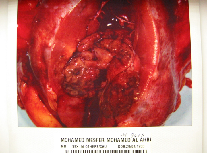
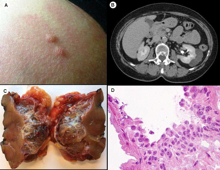
Introduction. Renal cell carcinoma is the third most common urogenital cancer. In some patients, it can metastasize to distant organs. Metastasis to the vagina is extremely rare. Case Presentation. A 54-year-old female with unremarkable history presented to the clinic with a chief complaint of vaginal bleeding. Further examination identified a pedunculated mass on the vaginal wall. Histologic examination revealed a metastatic clear cell renal cell carcinoma. Radiological studies then revealed a left renal mass and bilateral adrenal masses. The patient underwent a nephrectomy, adrenalectomy, and resection of the vaginal mass. The mass in the vagina has since recurred.We report the first known case of vaginal metastasis as initial presentation of a renal cell carcinoma with rhabdoid features. Postmenopausal women with renal cell carcinoma who present with vaginal bleeding should undergo a thorough inspection of the vaginal wall for the potential of metastatic neoplasms.
1. Introduction
2. Case Presentation
3. Discussion
4. Conclusion
Data Availability
Our conclusions arise from the evaluation of the histopathologic findings described in this study. No other data can be released due to patient confidentiality.
Conflicts of Interest
Don’t Miss: Basal Skin Cancer Survival Rates
Overview Of Origin Cell Type Stage And Grade
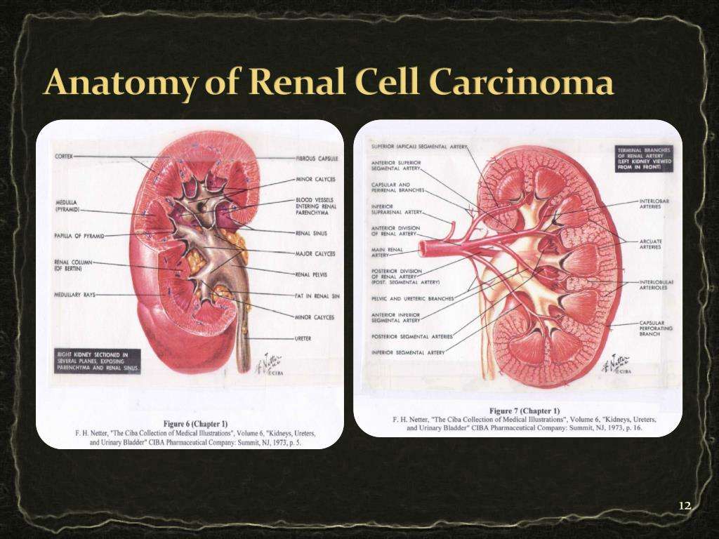
Human RCCs are thought to arise from a variety of specialized cells located along the length of the nephron. RCC is comprised of several histological cell types. Both clear cell and papillary RCC are thought to arise from the epithelium of the proximal tubule. Chromophobe RCC, oncocytoma, and collecting duct RCC are believed to arise from the distal nephron, probably from the epithelium of the collecting tubule. Each type has differences in genetics, biology and behavior. The most common histological type is clear cell carcinoma, also called conventional RCC, which represents 7580% of RCC. Papillary , chromophobe and other more rare forms such as collecting duct carcinoma comprise the remainder. Oncocytomas represent 37% of renal masses but are invariably benign and their exclusion from classification as RCC has been recommended . Distinct tumors of different cell types can occasionally be seen in the same kidney. An individual tumor can have mixed histologies. The pathologist differentiates cell types routinely by morphology and immunohistochemical markers as well as by cytogenetic and molecular genetic analysis particularly when the cell type is equivocal. Three to five per cent of RCC cannot be classified and are termed RCC, unclassified. Sarcomatoid RCC is no longer considered as a true subtype since sarcomatoid change represents undifferentiated cells associated with progression of disease in all RCC cell types .